5A7V

The GH130 family of mannoside phosphorylases contains glycoside hydrolases that target beta-1,2 mannosidic linkages in Candida mannan


Experimental Data Snapshot

  • Method: X-RAY DIFFRACTION
  • Resolution: 1.35 Å
  • R-Value Free: 0.150 
  • R-Value Work: 0.130 
  • R-Value Observed: 0.131 

wwPDB Validation   3D Report Full Report


Ligand Structure Quality Assessment 


This is version 1.4 of the entry. See complete history


Literature

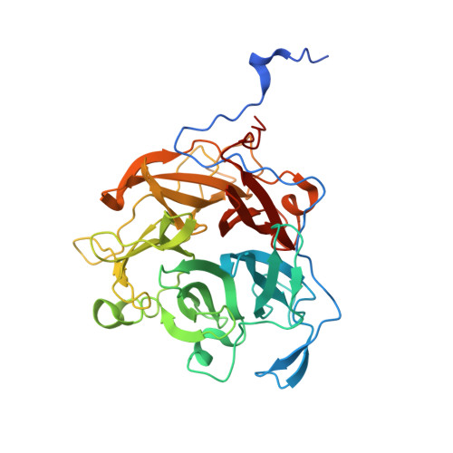
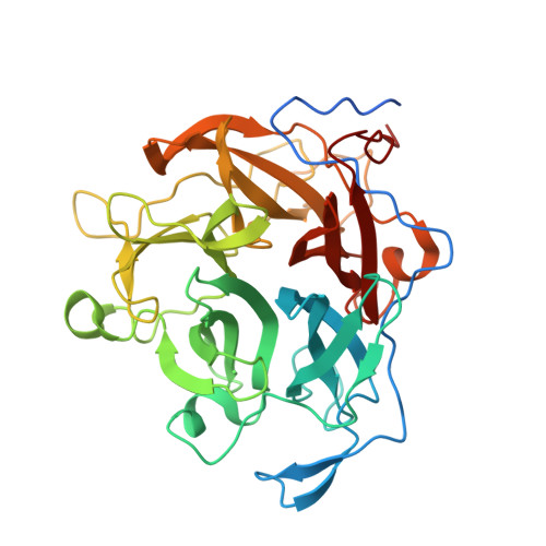
The Gh130 Family of Mannoside Phosphorylases Contains Glycoside Hydrolases that Target Beta-1,2 Mannosidic Linkages in Candida Mannan

Cuskin, F.Basle, A.Day, A.M.Ladeveze, S.Potocki-Veronese, G.Davies, G.J.Gilbert, H.J.Lowe, E.

(2015) J Biol Chem 290: 25023

  • DOI: https://doi.org/10.1074/jbc.M115.681460
  • Primary Citation of Related Structures:  
    5A7V

  • PubMed Abstract: 

    The depolymerization of complex glycans is an important biological process that is of considerable interest to environmentally relevant industries. β-Mannose is a major component of plant structural polysaccharides and eukaryotic N-glycans. These linkages are primarily cleaved by glycoside hydrolases, although recently, a family of glycoside phosphorylases, GH130, have also been shown to target β-1,2- and β-1,4-mannosidic linkages. In these phosphorylases, bond cleavage was mediated by a single displacement reaction in which phosphate functions as the catalytic nucleophile. A cohort of GH130 enzymes, however, lack the conserved basic residues that bind the phosphate nucleophile, and it was proposed that these enzymes function as glycoside hydrolases. Here we show that two Bacteroides enzymes, BT3780 and BACOVA_03624, which lack the phosphate binding residues, are indeed β-mannosidases that hydrolyze β-1,2-mannosidic linkages through an inverting mechanism. Because the genes encoding these enzymes are located in genetic loci that orchestrate the depolymerization of yeast α-mannans, it is likely that the two enzymes target the β-1,2-mannose residues that cap the glycan produced by Candida albicans. The crystal structure of BT3780 in complex with mannose bound in the -1 and +1 subsites showed that a pair of glutamates, Glu(227) and Glu(268), hydrogen bond to O1 of α-mannose, and either of these residues may function as the catalytic base. The candidate catalytic acid and the other residues that interact with the active site mannose are conserved in both GH130 mannoside phosphorylases and β-1,2-mannosidases. Functional phylogeny identified a conserved lysine, Lys(199) in BT3780, as a key specificity determinant for β-1,2-mannosidic linkages.


  • Organizational Affiliation

    From the Institute for Cell and Molecular Biosciences, Medical School Newcastle University, Newcastle upon Tyne NE2 4HH, United Kingdom.


Macromolecules
Find similar proteins by:  (by identity cutoff)  |  3D Structure
Entity ID: 1
MoleculeChains Sequence LengthOrganismDetailsImage
PUTATIVE GLYCOSIDASE PH117-RELATED388Bacteroides thetaiotaomicronMutation(s): 0 
UniProt
Find proteins for Q8A186 (Bacteroides thetaiotaomicron (strain ATCC 29148 / DSM 2079 / JCM 5827 / CCUG 10774 / NCTC 10582 / VPI-5482 / E50))
Explore Q8A186 
Go to UniProtKB:  Q8A186
Entity Groups  
Sequence Clusters30% Identity50% Identity70% Identity90% Identity95% Identity100% Identity
UniProt GroupQ8A186
Sequence Annotations
Expand
  • Reference Sequence
Find similar proteins by:  (by identity cutoff)  |  3D Structure
Entity ID: 2
MoleculeChains Sequence LengthOrganismDetailsImage
PUTATIVE GLYCOSIDASE PH117-RELATED388Bacteroides thetaiotaomicronMutation(s): 0 
UniProt
Find proteins for Q8A186 (Bacteroides thetaiotaomicron (strain ATCC 29148 / DSM 2079 / JCM 5827 / CCUG 10774 / NCTC 10582 / VPI-5482 / E50))
Explore Q8A186 
Go to UniProtKB:  Q8A186
Entity Groups  
Sequence Clusters30% Identity50% Identity70% Identity90% Identity95% Identity100% Identity
UniProt GroupQ8A186
Sequence Annotations
Expand
  • Reference Sequence
Small Molecules
Ligands 3 Unique
IDChains Name / Formula / InChI Key2D Diagram3D Interactions
MAN
Query on MAN

Download Ideal Coordinates CCD File 
D [auth A],
E [auth A],
M [auth B],
N [auth B]
alpha-D-mannopyranose
C6 H12 O6
WQZGKKKJIJFFOK-PQMKYFCFSA-N
BMA
Query on BMA

Download Ideal Coordinates CCD File 
C [auth A],
L [auth B]
beta-D-mannopyranose
C6 H12 O6
WQZGKKKJIJFFOK-RWOPYEJCSA-N
SO4
Query on SO4

Download Ideal Coordinates CCD File 
F [auth A]
G [auth A]
H [auth A]
I [auth A]
J [auth A]
F [auth A],
G [auth A],
H [auth A],
I [auth A],
J [auth A],
K [auth A],
O [auth B],
P [auth B],
Q [auth B],
R [auth B]
SULFATE ION
O4 S
QAOWNCQODCNURD-UHFFFAOYSA-L
Experimental Data & Validation

Experimental Data

  • Method: X-RAY DIFFRACTION
  • Resolution: 1.35 Å
  • R-Value Free: 0.150 
  • R-Value Work: 0.130 
  • R-Value Observed: 0.131 
  • Space Group: P 21 2 21
Unit Cell:
Length ( Å )Angle ( ˚ )
a = 75.767α = 90
b = 118.11β = 90
c = 126.586γ = 90
Software Package:
Software NamePurpose
REFMACrefinement
XDSdata reduction
Aimlessdata scaling
MOLREPphasing

Structure Validation

View Full Validation Report


Ligand Structure Quality Assessment 


Entry History 

Deposition Data

Revision History  (Full details and data files)

  • Version 1.0: 2015-08-26
    Type: Initial release
  • Version 1.1: 2015-09-02
    Changes: Database references
  • Version 1.2: 2015-10-21
    Changes: Database references
  • Version 1.3: 2020-07-29
    Type: Remediation
    Reason: Carbohydrate remediation
    Changes: Data collection, Derived calculations, Other, Structure summary
  • Version 1.4: 2024-01-10
    Changes: Data collection, Database references, Refinement description, Structure summary