


Structural and functional basis of difructose anhydride III hydrolase, which sequentially converts inulin using the same catalytic residue
Yu, S.H., Shen, H., Cheng, Y.Y., Zhu, Y.Y., Li, X., Mu, W.M.(2018) ACS Catal 8: 10683-10697
Experimental Data Snapshot
wwPDB Validation 3D Report Full Report

Entity ID: 1 | |||||
|---|---|---|---|---|---|
| Molecule | Chains | Sequence Length | Organism | Details | Image |
| DFA-IIIase | 445 | Pseudarthrobacter chlorophenolicus A6 | Mutation(s): 0 Gene Names: Achl_2895 EC: 3.2.1 | 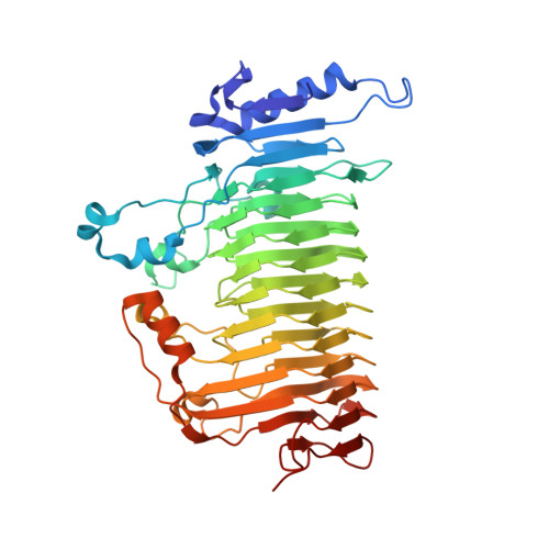 | |
UniProt | |||||
Find proteins for B8HDZ1 (Pseudarthrobacter chlorophenolicus (strain ATCC 700700 / DSM 12829 / CIP 107037 / JCM 12360 / KCTC 9906 / NCIMB 13794 / A6)) Explore B8HDZ1 Go to UniProtKB: B8HDZ1 | |||||
Entity Groups | |||||
| Sequence Clusters | 30% Identity50% Identity70% Identity90% Identity95% Identity100% Identity | ||||
| UniProt Group | B8HDZ1 | ||||
Sequence AnnotationsExpand | |||||
| |||||
| Length ( Å ) | Angle ( ˚ ) |
|---|---|
| a = 134.348 | α = 90 |
| b = 134.348 | β = 90 |
| c = 78.554 | γ = 120 |
| Software Name | Purpose |
|---|---|
| REFMAC | refinement |
| HKL-3000 | data reduction |
| HKL-3000 | data scaling |
| MOLREP | phasing |
RCSB PDB (citation) is hosted by
RCSB PDB is a member of the

