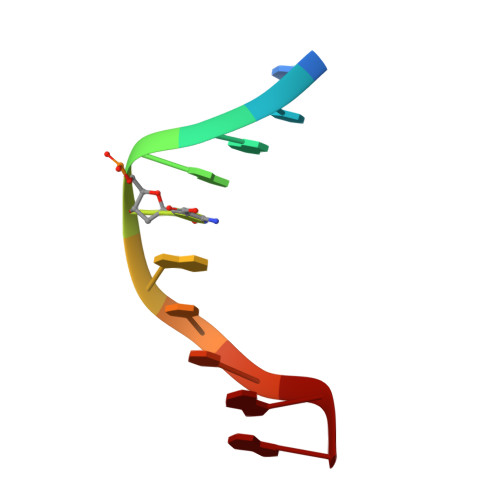
Thymine DNA glycosylase recognizes the geometry alteration of minor grooves induced by 5-formylcytosine and 5-carboxylcytosine.
Fu, T., Liu, L., Yang, Q.L., Wang, Y., Xu, P., Zhang, L., Liu, S., Dai, Q., Ji, Q., Xu, G.L., He, C., Luo, C., Zhang, L.(2019) Chem Sci 10: 7407-7417
- PubMed: 31489163
- DOI: https://doi.org/10.1039/c9sc02807b
- Primary Citation of Related Structures:
5ZAS, 5ZAT, 6JV3, 6JV5 - PubMed Abstract:
The dynamic DNA methylation-demethylation process plays critical roles in gene expression control and cell development. The oxidation derivatives of 5-methylcytosine (5mC) generated by Tet dioxygenases in the demethylation pathway, namely 5-hydroxymethylcytosine (5hmC), 5-formylcytosine (5fC), and 5-carboxylcytosine (5caC), could impact biological functions by altering DNA properties or recognition by potential reader proteins. Hence, in addition to the fifth base 5mC, 5hmC, 5fC, and 5caC have been considered as the sixth, seventh, and eighth bases of the genome. How these modifications would alter DNA and be specifically recognized remain unclear, however. Here we report that formyl- and carboxyl-modifications on cytosine induce the geometry alteration of the DNA minor groove by solving two high-resolution structures of a dsDNA decamer containing fully symmetric 5fC and 5caC. The alterations are recognized distinctively by thymine DNA glycosylase TDG via its finger residue R275, followed by subsequent preferential base excision and DNA repair. These observations suggest a mechanism by which reader proteins distinguish highly similar cytosine modifications for potential differential demethylation in order to achieve downstream biological functions.
Organizational Affiliation:
Department of Pharmacology and Chemical Biology , Shanghai Jiao Tong University School of Medicine , Shanghai , P. R. China . Email: liangzhang2014@sjtu.edu.cn ; Email: cluo@simm.ac.cn.


