5Y7M

Crystal structure of PhoRpp38 bound to a K-turn in P12.1 helix


Experimental Data Snapshot

  • Method: X-RAY DIFFRACTION
  • Resolution: 3.10 Å
  • R-Value Free: 0.247 
  • R-Value Work: 0.225 
  • R-Value Observed: 0.226 

Starting Model: experimental
View more details

wwPDB Validation   3D Report Full Report


Ligand Structure Quality Assessment 


This is version 1.3 of the entry. See complete history


Literature

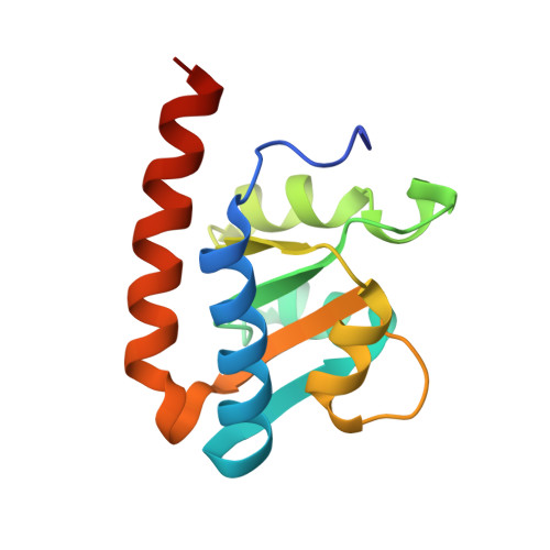
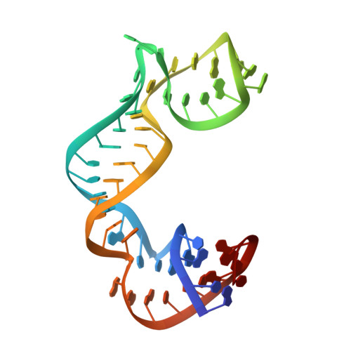
Crystal structures of the archaeal RNase P protein Rpp38 in complex with RNA fragments containing a K-turn motif.

Oshima, K.Gao, X.Hayashi, S.Ueda, T.Nakashima, T.Kimura, M.

(2018) Acta Crystallogr F Struct Biol Commun 74: 57-64

  • DOI: https://doi.org/10.1107/S2053230X17018039
  • Primary Citation of Related Structures:  
    5XTM, 5Y7M

  • PubMed Abstract: 

    A characteristic feature of archaeal ribonuclease P (RNase P) RNAs is that they have extended helices P12.1 and P12.2 containing kink-turn (K-turn) motifs to which the archaeal RNase P protein Rpp38, a homologue of the human RNase P protein Rpp38, specifically binds. PhoRpp38 from the hyperthermophilic archaeon Pyrococcus horikoshii is involved in the elevation of the optimum temperature of the reconstituted RNase P by binding the K-turns in P12.1 and P12.2. Previously, the crystal structure of PhoRpp38 in complex with the K-turn in P12.2 was determined at 3.4 Å resolution. In this study, the crystal structure of PhoRpp38 in complex with the K-turn in P12.2 was improved to 2.1 Å resolution and the structure of PhoRpp38 in complex with the K-turn in P12.1 was also determined at a resolution of 3.1 Å. Both structures revealed that Lys35, Asn38 and Glu39 in PhoRpp38 interact with characteristic G·A and A·G pairs in the K-turn, while Thr37, Asp59, Lys84, Glu94, Ala96 and Ala98 in PhoRpp38 interact with the three-nucleotide bulge in the K-turn. Moreover, an extended stem-loop containing P10-P12.2 in complex with PhoRpp38, as well as PhoRpp21 and PhoRpp29, which are the archaeal homologues of the human proteins Rpp21 and Rpp29, respectively, was affinity-purified and crystallized. The crystals thus grown diffracted to a resolution of 6.35 Å. Structure determination of the crystals will demonstrate the previously proposed secondary structure of stem-loops including helices P12.1 and P12.2 and will also provide insight into the structural organization of the specificity domain in P. horikoshii RNase P RNA.


  • Organizational Affiliation

    Laboratory of Biochemistry, Department of Bioscience and Biotechnology, Graduate School, Faculty of Agriculture, Kyushu University, Hakozaki 6-10-1, Fukuoka 812-8581, Japan.


Macromolecules

Find similar proteins by:  (by identity cutoff)  |  3D Structure
Entity ID: 1
MoleculeChains Sequence LengthOrganismDetailsImage
50S ribosomal protein L7Ae
A, C
132Pyrococcus horikoshii OT3Mutation(s): 0 
Gene Names: rpl7aePH1496
UniProt
Find proteins for P62009 (Pyrococcus horikoshii (strain ATCC 700860 / DSM 12428 / JCM 9974 / NBRC 100139 / OT-3))
Explore P62009 
Go to UniProtKB:  P62009
Entity Groups  
Sequence Clusters30% Identity50% Identity70% Identity90% Identity95% Identity100% Identity
UniProt GroupP62009
Sequence Annotations
Expand
  • Reference Sequence
Find similar nucleic acids by:  (by identity cutoff)  |  3D Structure
Entity ID: 2
MoleculeChains LengthOrganismImage
RNA (52-MER)
B, D
51Pyrococcus horikoshii OT3
Sequence Annotations
Expand
  • Reference Sequence
Small Molecules
Ligands 2 Unique
IDChains Name / Formula / InChI Key2D Diagram3D Interactions
GTP
Query on GTP

Download Ideal Coordinates CCD File 
E [auth B],
L [auth D]
GUANOSINE-5'-TRIPHOSPHATE
C10 H16 N5 O14 P3
XKMLYUALXHKNFT-UUOKFMHZSA-N
SO4
Query on SO4

Download Ideal Coordinates CCD File 
F [auth B]
G [auth B]
H [auth B]
I [auth C]
J [auth C]
F [auth B],
G [auth B],
H [auth B],
I [auth C],
J [auth C],
K [auth C],
M [auth D],
N [auth D],
O [auth D]
SULFATE ION
O4 S
QAOWNCQODCNURD-UHFFFAOYSA-L
Experimental Data & Validation

Experimental Data

  • Method: X-RAY DIFFRACTION
  • Resolution: 3.10 Å
  • R-Value Free: 0.247 
  • R-Value Work: 0.225 
  • R-Value Observed: 0.226 
  • Space Group: C 1 2 1
Unit Cell:
Length ( Å )Angle ( ˚ )
a = 113.37α = 90
b = 176.6β = 105.12
c = 59.9γ = 90
Software Package:
Software NamePurpose
PHENIXrefinement
XSCALEdata scaling
PHASERphasing
PDB_EXTRACTdata extraction

Structure Validation

View Full Validation Report


Ligand Structure Quality Assessment 


Entry History 

Deposition Data

Revision History  (Full details and data files)

  • Version 1.0: 2018-02-07
    Type: Initial release
  • Version 1.1: 2020-02-26
    Changes: Data collection
  • Version 1.2: 2023-11-22
    Changes: Data collection, Database references, Refinement description
  • Version 1.3: 2024-03-20
    Changes: Source and taxonomy