


Crystal structure of SmyD3 in complex with covalent inhibitor 2
Baburajendran, N., Jansson, E.A.To be published.
Experimental Data Snapshot
Entity ID: 1 | |||||
|---|---|---|---|---|---|
| Molecule | Chains | Sequence Length | Organism | Details | Image |
| SMYD3 | 423 | Homo sapiens | Mutation(s): 0 | 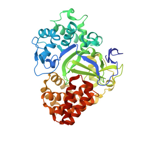 | |
UniProt & NIH Common Fund Data Resources | |||||
Find proteins for Q9H7B4 (Homo sapiens) Explore Q9H7B4 Go to UniProtKB: Q9H7B4 | |||||
PHAROS: Q9H7B4 GTEx: ENSG00000185420 | |||||
Entity Groups | |||||
| Sequence Clusters | 30% Identity50% Identity70% Identity90% Identity95% Identity100% Identity | ||||
| UniProt Group | Q9H7B4 | ||||
Sequence AnnotationsExpand | |||||
| |||||
| Ligands 3 Unique | |||||
|---|---|---|---|---|---|
| ID | Chains | Name / Formula / InChI Key | 2D Diagram | 3D Interactions | |
| 8HR Query on 8HR | B [auth A] | propyl 4-(4-chloranyl-2-phenyl-quinolin-7-yl)carbonylpiperazine-1-carboxylate C24 H24 Cl N3 O3 KSURKAFQFJOCBC-UHFFFAOYSA-N |  | ||
| SAM Query on SAM | C [auth A] | S-ADENOSYLMETHIONINE C15 H22 N6 O5 S MEFKEPWMEQBLKI-FCKMPRQPSA-N |  | ||
| ZN Query on ZN | D [auth A], E [auth A], F [auth A] | ZINC ION Zn PTFCDOFLOPIGGS-UHFFFAOYSA-N |  | ||
| Length ( Å ) | Angle ( ˚ ) |
|---|---|
| a = 61.288 | α = 90 |
| b = 66.374 | β = 90 |
| c = 107.296 | γ = 90 |
| Software Name | Purpose |
|---|---|
| HKL-2000 | data scaling |
| PHENIX | refinement |
| PDB_EXTRACT | data extraction |
RCSB PDB (citation) is hosted by
RCSB PDB is a member of the

