5XUJ

Crystal structure of PDE10A in complex with 7-(4-chlorophenyl)-2-methylpyrazolo[1,5-a]pyrimidine


Experimental Data Snapshot

  • Method: X-RAY DIFFRACTION
  • Resolution: 2.44 Å
  • R-Value Free: 0.280 
  • R-Value Work: 0.198 
  • R-Value Observed: 0.202 

wwPDB Validation   3D Report Full Report


Ligand Structure Quality Assessment 


This is version 1.1 of the entry. See complete history


Literature

Fragment-Based Discovery of Pyrimido[1,2-b]indazole PDE10A Inhibitors.

Chino, A.Seo, R.Amano, Y.Namatame, I.Hamaguchi, W.Honbou, K.Mihara, T.Yamazaki, M.Tomishima, M.Masuda, N.

(2018) Chem Pharm Bull (Tokyo) 66: 286-294

  • DOI: https://doi.org/10.1248/cpb.c17-00836
  • Primary Citation of Related Structures:  
    5XUI, 5XUJ

  • PubMed Abstract: 

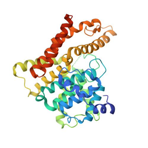
    In this study, we report the identification of potent pyrimidoindazoles as phosphodiesterase10A (PDE10A) inhibitors by using the method of fragment-based drug discovery (FBDD). The pyrazolopyridine derivative 2 was found to be a fragment hit compound which could occupy a part of the binding site of PDE10A enzyme by using the method of the X-ray co-crystal structure analysis. On the basis of the crystal structure of compound 2 and PDE10A protein, a number of compounds were synthesized and evaluated, by means of structure-activity relationship (SAR) studies, which culminated in the discovery of a novel pyrimidoindazole derivative 13 having good physicochemical properties.


  • Organizational Affiliation

    Drug Discovery Research, Astellas Pharma Inc.


Macromolecules
Find similar proteins by:  (by identity cutoff)  |  3D Structure
Entity ID: 1
MoleculeChains Sequence LengthOrganismDetailsImage
cAMP and cAMP-inhibited cGMP 3',5'-cyclic phosphodiesterase 10A
A, B
345Homo sapiensMutation(s): 0 
Gene Names: PDE10A
EC: 3.1.4.17 (PDB Primary Data), 3.1.4.35 (PDB Primary Data)
UniProt & NIH Common Fund Data Resources
Find proteins for Q9Y233 (Homo sapiens)
Explore Q9Y233 
Go to UniProtKB:  Q9Y233
PHAROS:  Q9Y233
GTEx:  ENSG00000112541 
Entity Groups  
Sequence Clusters30% Identity50% Identity70% Identity90% Identity95% Identity100% Identity
UniProt GroupQ9Y233
Sequence Annotations
Expand
  • Reference Sequence
Experimental Data & Validation

Experimental Data

  • Method: X-RAY DIFFRACTION
  • Resolution: 2.44 Å
  • R-Value Free: 0.280 
  • R-Value Work: 0.198 
  • R-Value Observed: 0.202 
  • Space Group: P 21 21 21
Unit Cell:
Length ( Å )Angle ( ˚ )
a = 50.339α = 90
b = 81.299β = 90
c = 157.597γ = 90
Software Package:
Software NamePurpose
REFMACrefinement
HKL-2000data reduction
HKL-2000data scaling
AMoREphasing

Structure Validation

View Full Validation Report


Ligand Structure Quality Assessment 


Entry History 

Deposition Data

Revision History  (Full details and data files)

  • Version 1.0: 2018-03-14
    Type: Initial release
  • Version 1.1: 2024-03-27
    Changes: Data collection, Database references, Derived calculations