5XIE

Crystal Structure of HasAp with 5-ethynyl-10,20-diphenylporphyrin


Experimental Data Snapshot

  • Method: X-RAY DIFFRACTION
  • Resolution: 2.05 Å
  • R-Value Free: 0.203 
  • R-Value Work: 0.173 
  • R-Value Observed: 0.174 

wwPDB Validation   3D Report Full Report


Ligand Structure Quality Assessment 


This is version 1.1 of the entry. See complete history


Literature

Structures of the Heme Acquisition Protein HasA with Iron(III)-5,15-Diphenylporphyrin and Derivatives Thereof as an Artificial Prosthetic Group

Uehara, H.Shisaka, Y.Nishimura, T.Sugimoto, H.Shiro, Y.Miyake, Y.Shinokubo, H.Watanabe, Y.Shoji, O.

(2017) Angew Chem Int Ed Engl 56: 15279-15283

  • DOI: https://doi.org/10.1002/anie.201707212
  • Primary Citation of Related Structures:  
    5XA4, 5XIB, 5XIC, 5XIE, 5XKB

  • PubMed Abstract: 

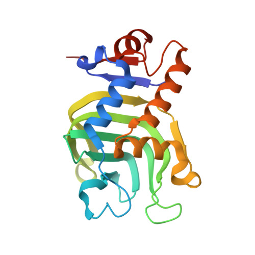
    Iron(III)-5,15-diphenylporphyrin and several derivatives were accommodated by HasA, a heme acquisition protein secreted by Pseudomonas aeruginosa, despite possessing bulky substituents at the meso position of the porphyrin. Crystal structure analysis revealed that the two phenyl groups at the meso positions of porphyrin extend outside HasA. It was shown that the growth of P. aeruginosa was inhibited in the presence of HasA coordinating the synthetic porphyrins under iron-limiting conditions, and that the structure of the synthetic porphyrins greatly affects the inhibition efficiency.


  • Organizational Affiliation

    Department of Chemistry, Graduate School of Science, Nagoya University, Furo-cho, Chikusa-ku, Nagoya, 464-8602, Japan.


Macromolecules
Find similar proteins by:  (by identity cutoff)  |  3D Structure
Entity ID: 1
MoleculeChains Sequence LengthOrganismDetailsImage
Heme acquisition protein HasAp
A, B
184Pseudomonas aeruginosa PAO1Mutation(s): 0 
Gene Names: hasApPA3407
UniProt
Find proteins for G3XD33 (Pseudomonas aeruginosa (strain ATCC 15692 / DSM 22644 / CIP 104116 / JCM 14847 / LMG 12228 / 1C / PRS 101 / PAO1))
Explore G3XD33 
Go to UniProtKB:  G3XD33
Entity Groups  
Sequence Clusters30% Identity50% Identity70% Identity90% Identity95% Identity100% Identity
UniProt GroupG3XD33
Sequence Annotations
Expand
  • Reference Sequence
Small Molecules
Ligands 1 Unique
IDChains Name / Formula / InChI Key2D Diagram3D Interactions
WYP
Query on WYP

Download Ideal Coordinates CCD File 
C [auth A],
D [auth B]
5-Ethynyl-10,20-diphenylporphyrin containing FE
C34 H20 Fe N4
REUZHWAQZCOEAH-GGYHVSAHSA-N
Experimental Data & Validation

Experimental Data

  • Method: X-RAY DIFFRACTION
  • Resolution: 2.05 Å
  • R-Value Free: 0.203 
  • R-Value Work: 0.173 
  • R-Value Observed: 0.174 
  • Space Group: P 61
Unit Cell:
Length ( Å )Angle ( ˚ )
a = 153.622α = 90
b = 153.622β = 90
c = 36.825γ = 120
Software Package:
Software NamePurpose
REFMACrefinement
DENZOdata collection
SCALEPACKdata scaling
PDB_EXTRACTdata extraction
DENZOdata reduction
MOLREPphasing

Structure Validation

View Full Validation Report


Ligand Structure Quality Assessment 


Entry History 

Deposition Data

Revision History  (Full details and data files)

  • Version 1.0: 2017-12-06
    Type: Initial release
  • Version 1.1: 2023-11-22
    Changes: Data collection, Database references, Refinement description