


Crystal structure of VqsR LBD domain from Pseudomonas aeruginosa
Gu, L., He, Q., Wang, K., Wang, F.To be published.
Experimental Data Snapshot
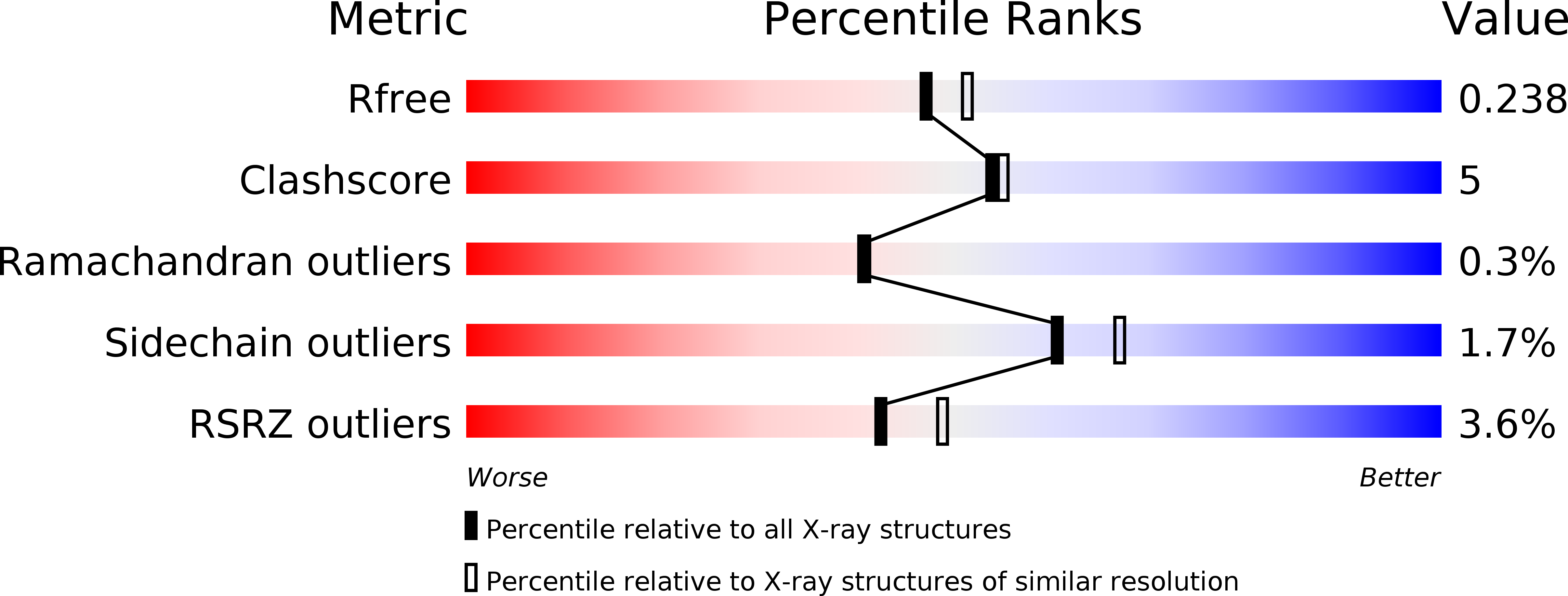
wwPDB Validation 3D Report Full Report
Entity ID: 1 | |||||
|---|---|---|---|---|---|
| Molecule | Chains | Sequence Length | Organism | Details | Image |
| Helix-turn-helix transcriptional regulator | 195 | Pseudomonas aeruginosa | Mutation(s): 0 Gene Names: PA2591, nreC_2, nreC, nreC_1, nreC_3, AOY09_03830, BH593_21715, CCBH4851_00420, PAERUG_E15_London_28_01_14_06660, PAERUG_P32_London_17_VIM_2_10_11_00166... | 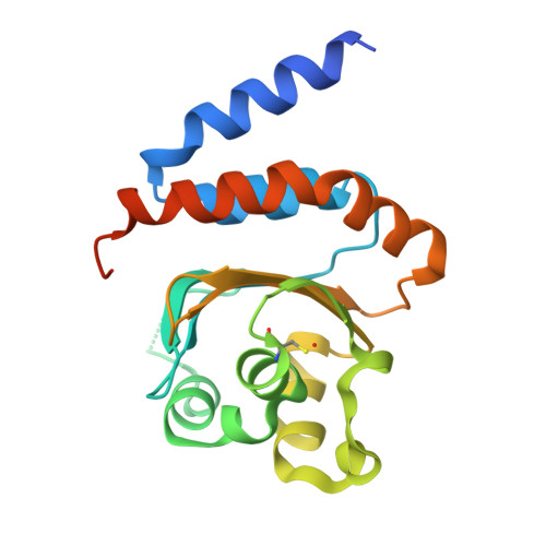 | |
UniProt | |||||
Find proteins for Q9I0P6 (Pseudomonas aeruginosa (strain ATCC 15692 / DSM 22644 / CIP 104116 / JCM 14847 / LMG 12228 / 1C / PRS 101 / PAO1)) Explore Q9I0P6 Go to UniProtKB: Q9I0P6 | |||||
Entity Groups | |||||
| Sequence Clusters | 30% Identity50% Identity70% Identity90% Identity95% Identity100% Identity | ||||
| UniProt Group | Q9I0P6 | ||||
Sequence AnnotationsExpand | |||||
| |||||
| Modified Residues 1 Unique | |||||
|---|---|---|---|---|---|
| ID | Chains | Type | Formula | 2D Diagram | Parent |
| CSO Query on CSO | A, B | L-PEPTIDE LINKING | C3 H7 N O3 S |  | CYS |
| Length ( Å ) | Angle ( ˚ ) |
|---|---|
| a = 52.834 | α = 90 |
| b = 135.198 | β = 90 |
| c = 150.772 | γ = 90 |
| Software Name | Purpose |
|---|---|
| PHENIX | refinement |
| HKL-2000 | data reduction |
| HKL-2000 | data scaling |
| PHENIX | phasing |
RCSB PDB (citation) is hosted by
RCSB PDB is a member of the

