


Crystal Structure of HasAp with Gallium Phthalocyanine
Shoji, O., Shisaka, Y., Iwai, Y., Sugimoto, H., Shiro, Y., Watanabe, Y.To be published.
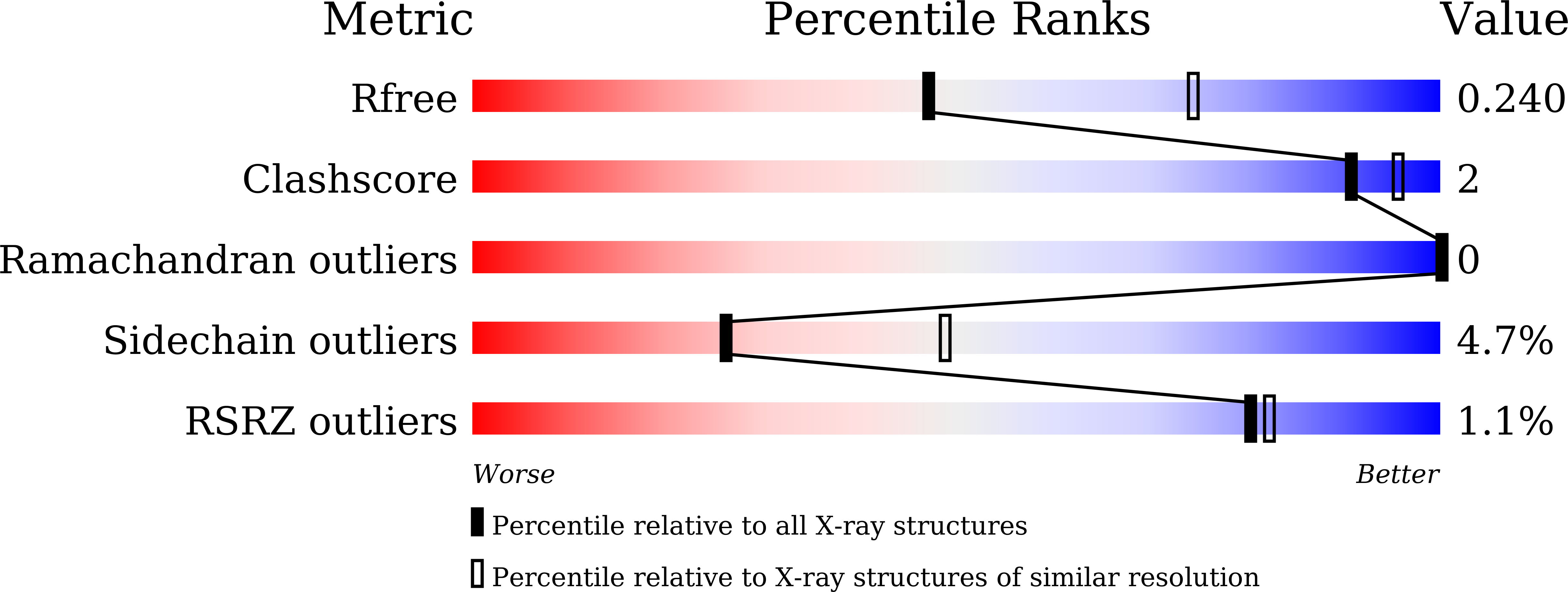
Experimental Data Snapshot
Entity ID: 1 | |||||
|---|---|---|---|---|---|
| Molecule | Chains | Sequence Length | Organism | Details | Image |
| Heme acquisition protein HasAp | 184 | Pseudomonas aeruginosa PAO1 | Mutation(s): 0 Gene Names: hasAp, PA3407 | 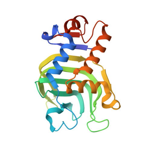 | |
UniProt | |||||
Find proteins for G3XD33 (Pseudomonas aeruginosa (strain ATCC 15692 / DSM 22644 / CIP 104116 / JCM 14847 / LMG 12228 / 1C / PRS 101 / PAO1)) Explore G3XD33 Go to UniProtKB: G3XD33 | |||||
Entity Groups | |||||
| Sequence Clusters | 30% Identity50% Identity70% Identity90% Identity95% Identity100% Identity | ||||
| UniProt Group | G3XD33 | ||||
Sequence AnnotationsExpand | |||||
| |||||
| Ligands 1 Unique | |||||
|---|---|---|---|---|---|
| ID | Chains | Name / Formula / InChI Key | 2D Diagram | 3D Interactions | |
| VPC Query on VPC | C [auth A], D [auth B] | Phthalocyanine containing GA C32 H16 Ga N8 WBSOKGMOXCALAG-UHFFFAOYSA-N |  | ||
| Length ( Å ) | Angle ( ˚ ) |
|---|---|
| a = 153.625 | α = 90 |
| b = 153.625 | β = 90 |
| c = 36.761 | γ = 120 |
| Software Name | Purpose |
|---|---|
| REFMAC | refinement |
| DENZO | data collection |
| SCALEPACK | data scaling |
| PDB_EXTRACT | data extraction |
| DENZO | data reduction |
| MOLREP | phasing |
RCSB PDB (citation) is hosted by
RCSB PDB is a member of the

