5XBF

Crystal Structure of Myo7b C-terminal MyTH4-FERM in complex with USH1C PDZ3


Experimental Data Snapshot

  • Method: X-RAY DIFFRACTION
  • Resolution: 1.80 Å
  • R-Value Free: 0.211 
  • R-Value Work: 0.187 
  • R-Value Observed: 0.188 

wwPDB Validation   3D Report Full Report


This is version 1.2 of the entry. See complete history


Literature

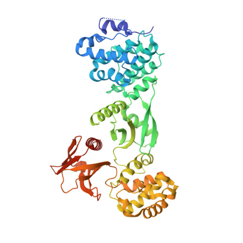
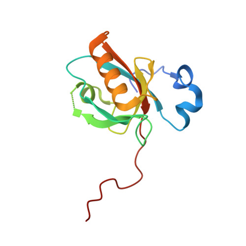
Structure of Myo7b/USH1C complex suggests a general PDZ domain binding mode by MyTH4-FERM myosins.

Li, J.He, Y.Weck, M.L.Lu, Q.Tyska, M.J.Zhang, M.

(2017) Proc Natl Acad Sci U S A 114: E3776-E3785

  • DOI: https://doi.org/10.1073/pnas.1702251114
  • Primary Citation of Related Structures:  
    5XBF

  • PubMed Abstract: 

    Unconventional myosin 7a (Myo7a), myosin 7b (Myo7b), and myosin 15a (Myo15a) all contain MyTH4-FERM domains (myosin tail homology 4-band 4.1, ezrin, radixin, moesin; MF) in their cargo binding tails and are essential for the growth and function of microvilli and stereocilia. Numerous mutations have been identified in the MyTH4-FERM tandems of these myosins in patients suffering visual and hearing impairment. Although a number of MF domain binding partners have been identified, the molecular basis of interactions with the C-terminal MF domain (CMF) of these myosins remains poorly understood. Here we report the high-resolution crystal structure of Myo7b CMF in complex with the extended PDZ3 domain of USH1C (a.k.a., Harmonin), revealing a previously uncharacterized interaction mode both for MyTH4-FERM tandems and for PDZ domains. We predicted, based on the structure of the Myo7b CMF/USH1C PDZ3 complex, and verified that Myo7a CMF also binds to USH1C PDZ3 using a similar mode. The structure of the Myo7b CMF/USH1C PDZ complex provides mechanistic explanations for >20 deafness-causing mutations in Myo7a CMF. Taken together, these findings suggest that binding to PDZ domains, such as those from USH1C, PDZD7, and Whirlin, is a common property of CMFs of Myo7a, Myo7b, and Myo15a.


  • Organizational Affiliation

    Division of Life Science, State Key Laboratory of Molecular Neuroscience, Hong Kong University of Science and Technology, Clear Water Bay, Kowloon, Hong Kong.


Macromolecules
Find similar proteins by:  (by identity cutoff)  |  3D Structure
Entity ID: 1
MoleculeChains Sequence LengthOrganismDetailsImage
Unconventional myosin-VIIb516Homo sapiensMutation(s): 0 
Gene Names: MYO7B
UniProt & NIH Common Fund Data Resources
Find proteins for Q6PIF6 (Homo sapiens)
Explore Q6PIF6 
Go to UniProtKB:  Q6PIF6
GTEx:  ENSG00000169994 
Entity Groups  
Sequence Clusters30% Identity50% Identity70% Identity90% Identity95% Identity100% Identity
UniProt GroupQ6PIF6
Sequence Annotations
Expand
  • Reference Sequence
Find similar proteins by:  (by identity cutoff)  |  3D Structure
Entity ID: 2
MoleculeChains Sequence LengthOrganismDetailsImage
Harmonin125Homo sapiensMutation(s): 0 
Gene Names: USH1CAIE75
UniProt & NIH Common Fund Data Resources
Find proteins for Q9Y6N9 (Homo sapiens)
Explore Q9Y6N9 
Go to UniProtKB:  Q9Y6N9
PHAROS:  Q9Y6N9
GTEx:  ENSG00000006611 
Entity Groups  
Sequence Clusters30% Identity50% Identity70% Identity90% Identity95% Identity100% Identity
UniProt GroupQ9Y6N9
Sequence Annotations
Expand
  • Reference Sequence
Experimental Data & Validation

Experimental Data

  • Method: X-RAY DIFFRACTION
  • Resolution: 1.80 Å
  • R-Value Free: 0.211 
  • R-Value Work: 0.187 
  • R-Value Observed: 0.188 
  • Space Group: P 1 21 1
Unit Cell:
Length ( Å )Angle ( ˚ )
a = 69.634α = 90
b = 42.537β = 97.51
c = 118.39γ = 90
Software Package:
Software NamePurpose
DENZOdata collection
SCALEPACKdata scaling
PHENIXrefinement
PDB_EXTRACTdata extraction
DENZOdata reduction
AutoSolphasing

Structure Validation

View Full Validation Report


Entry History 

Deposition Data

Revision History  (Full details and data files)

  • Version 1.0: 2017-05-17
    Type: Initial release
  • Version 1.1: 2017-12-06
    Changes: Database references
  • Version 1.2: 2024-03-27
    Changes: Data collection, Database references