Crystal Structure and Substrate Specificity Modification of Acetyl Xylan Esterase from Aspergillus luchuensis
Komiya, D., Hori, A., Ishida, T., Igarashi, K., Samejima, M., Koseki, T., Fushinobu, S.(2017) Appl Environ Microbiol 83
- PubMed: 28802264
- DOI: https://doi.org/10.1128/AEM.01251-17
- Primary Citation of Related Structures:
5X6S - PubMed Abstract:
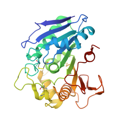
Acetyl xylan esterase (AXE) catalyzes the hydrolysis of the acetyl bonds present in plant cell wall polysaccharides. Here, we determined the crystal structure of AXE from Aspergillus luchuensis ( Al AXEA), providing the three-dimensional structure of an enzyme in the Esterase_phb family. Al AXEA shares its core α/β-hydrolase fold structure with esterases in other families, but it has an extended central β-sheet at both its ends and an extra loop. Structural comparison with a ferulic acid esterase (FAE) from Aspergillus niger indicated that Al AXEA has a conserved catalytic machinery: a catalytic triad (Ser119, His259, and Asp202) and an oxyanion hole (Cys40 and Ser120). Near the catalytic triad of A lAXEA, two aromatic residues (Tyr39 and Trp160) form small pockets at both sides. Homology models of fungal FAEs in the same Esterase_phb family have wide pockets at the corresponding sites because they have residues with smaller side chains (Pro, Ser, and Gly). Mutants with site-directed mutations at Tyr39 showed a substrate specificity similar to that of the wild-type enzyme, whereas those with mutations at Trp160 acquired an expanded substrate specificity. Interestingly, the Trp160 mutants acquired weak but significant type B-like FAE activity. Moreover, the engineered enzymes exhibited ferulic acid-releasing activity from wheat arabinoxylan. IMPORTANCE Hemicelluloses in the plant cell wall are often decorated by acetyl and ferulic acid groups. Therefore, complete and efficient degradation of plant polysaccharides requires the enzymes for cleaving the side chains of the polymer. Since the Esterase_phb family contains a wide array of fungal FAEs and AXEs from fungi and bacteria, our study will provide a structural basis for the molecular mechanism of these industrially relevant enzymes in biopolymer degradation. The structure of the Esterase_phb family also provides information for bacterial polyhydroxyalkanoate depolymerases that are involved in biodegradation of thermoplastic polymers.
Organizational Affiliation:
Department of Biotechnology, The University of Tokyo, Tokyo, Japan.