5WUN

Crystal structure of mouse importin-alpha1 bound to non-phosphorylated NLS of EBNA1


Experimental Data Snapshot

  • Method: X-RAY DIFFRACTION
  • Resolution: 2.20 Å
  • R-Value Free: 0.204 
  • R-Value Work: 0.177 
  • R-Value Observed: 0.178 

wwPDB Validation   3D Report Full Report


This is version 1.2 of the entry. See complete history


Literature

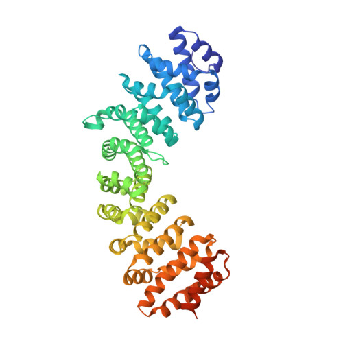
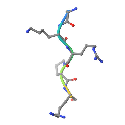
Structural basis for the regulation of nuclear import of Epstein-Barr virus nuclear antigen 1 (EBNA1) by phosphorylation of the nuclear localization signal.

Nakada, R.Hirano, H.Matsuura, Y.

(2017) Biochem Biophys Res Commun 484: 113-117

  • DOI: https://doi.org/10.1016/j.bbrc.2017.01.063
  • Primary Citation of Related Structures:  
    5WUM, 5WUN

  • PubMed Abstract: 

    Epstein-Barr virus (EBV) nuclear antigen 1 (EBNA1) is expressed in every EBV-positive tumor and is essential for the maintenance, replication, and transcription of the EBV genome in the nucleus of host cells. EBNA1 is a serine phosphoprotein, and it has been shown that phosphorylation of S385 in the nuclear localization signal (NLS) of EBNA1 increases the binding affinity to the nuclear import adaptor importin-α1 as well as importin-α5, and stimulates nuclear import of EBNA1. To gain insights into how phosphorylation of the EBNA1 NLS regulates nuclear import, we have determined the crystal structures of two peptide complexes of importin-α1: one with S385-phosphorylated EBNA1 NLS peptide, determined at 2.0 Å resolution, and one with non-phosphorylated EBNA1 NLS peptide, determined at 2.2 Å resolution. The structures show that EBNA1 NLS binds to the major and minor NLS-binding sites of importin-α1, and indicate that the binding affinity of the EBNA1 NLS to the minor NLS-binding site could be enhanced by phosphorylation of S385 through electrostatic interaction between the phosphate group of phospho-S385 and K392 of importin-α1 (corresponding to R395 of importin-α5) on armadillo repeat 8.


  • Organizational Affiliation

    Division of Biological Science, Graduate School of Science, Nagoya University, Japan.


Macromolecules
Find similar proteins by:  (by identity cutoff)  |  3D Structure
Entity ID: 1
MoleculeChains Sequence LengthOrganismDetailsImage
Importin subunit alpha-1460Mus musculusMutation(s): 0 
Gene Names: Kpna2Rch1
UniProt & NIH Common Fund Data Resources
Find proteins for P52293 (Mus musculus)
Explore P52293 
Go to UniProtKB:  P52293
IMPC:  MGI:103561
Entity Groups  
Sequence Clusters30% Identity50% Identity70% Identity90% Identity95% Identity100% Identity
UniProt GroupP52293
Sequence Annotations
Expand
  • Reference Sequence

Find similar proteins by:  Sequence   |   3D Structure  

Entity ID: 2
MoleculeChains Sequence LengthOrganismDetailsImage
Epstein-Barr nuclear antigen 1
B, C
9Human herpesvirus 4 strain B95-8Mutation(s): 0 
UniProt
Find proteins for P03211 (Epstein-Barr virus (strain B95-8))
Explore P03211 
Go to UniProtKB:  P03211
Entity Groups  
UniProt GroupP03211
Sequence Annotations
Expand
  • Reference Sequence
Experimental Data & Validation

Experimental Data

  • Method: X-RAY DIFFRACTION
  • Resolution: 2.20 Å
  • R-Value Free: 0.204 
  • R-Value Work: 0.177 
  • R-Value Observed: 0.178 
  • Space Group: P 21 21 21
Unit Cell:
Length ( Å )Angle ( ˚ )
a = 78.51α = 90
b = 90.18β = 90
c = 98.89γ = 90
Software Package:
Software NamePurpose
MOSFLMdata collection
Aimlessdata scaling
MOLREPphasing
PHENIXrefinement
PDB_EXTRACTdata extraction
MOSFLMdata reduction

Structure Validation

View Full Validation Report


Entry History 

Deposition Data

Revision History  (Full details and data files)

  • Version 1.0: 2017-01-25
    Type: Initial release
  • Version 1.1: 2017-12-06
    Changes: Data collection, Database references
  • Version 1.2: 2023-11-22
    Changes: Data collection, Database references, Refinement description