


Expression, Biochemical Characterization and Structure Resolution of beta-glucosidase from Paenibacillus barengoltzii
Huang, P., Wu, S., Jiang, Z., Yang, S.(2019) J Food Sci Technol(china)
Experimental Data Snapshot
wwPDB Validation 3D Report Full Report

Entity ID: 1 | |||||
|---|---|---|---|---|---|
| Molecule | Chains | Sequence Length | Organism | Details | Image |
| Beta-glucosidase | 955 | Paenibacillus barengoltzii | Mutation(s): 0 EC: 3.2.1.21 | 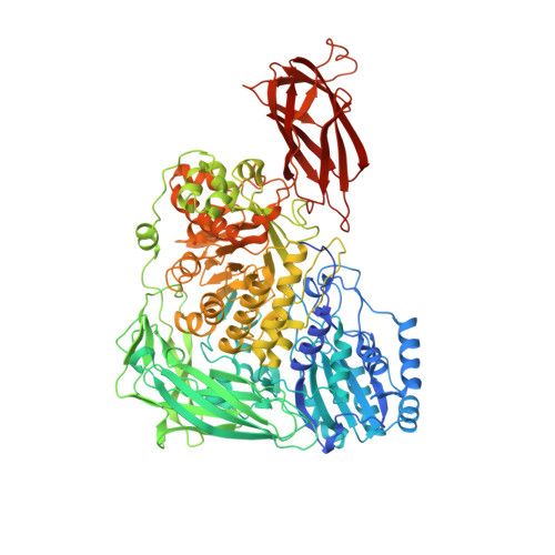 | |
UniProt | |||||
Find proteins for A0A1P8VKA0 (Paenibacillus barengoltzii) Explore A0A1P8VKA0 Go to UniProtKB: A0A1P8VKA0 | |||||
Entity Groups | |||||
| Sequence Clusters | 30% Identity50% Identity70% Identity90% Identity95% Identity100% Identity | ||||
| UniProt Group | A0A1P8VKA0 | ||||
Sequence AnnotationsExpand | |||||
| |||||
| Length ( Å ) | Angle ( ˚ ) |
|---|---|
| a = 67.219 | α = 90 |
| b = 75.786 | β = 90 |
| c = 161.994 | γ = 90 |
| Software Name | Purpose |
|---|---|
| PHENIX | refinement |
| HKL-2000 | data processing |
| HKL-2000 | data scaling |
| PHENIX | model building |
RCSB PDB (citation) is hosted by
RCSB PDB is a member of the

