


Crystal structure of NAD synthetase NadE from Vibrio fischeri
Stogios, P.J.To be published.
Experimental Data Snapshot
wwPDB Validation 3D Report Full Report

Entity ID: 1 | |||||
|---|---|---|---|---|---|
| Molecule | Chains | Sequence Length | Organism | Details | Image |
| NH(3)-dependent NAD(+) synthetase | 279 | Aliivibrio fischeri ES114 | Mutation(s): 0 Gene Names: nadE, VF_A0602 EC: 6.3.1.5 | 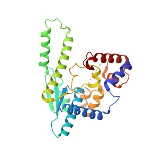 | |
UniProt | |||||
Find proteins for Q5DZX4 (Aliivibrio fischeri (strain ATCC 700601 / ES114)) Explore Q5DZX4 Go to UniProtKB: Q5DZX4 | |||||
Entity Groups | |||||
| Sequence Clusters | 30% Identity50% Identity70% Identity90% Identity95% Identity100% Identity | ||||
| UniProt Group | Q5DZX4 | ||||
Sequence AnnotationsExpand | |||||
| |||||
| Length ( Å ) | Angle ( ˚ ) |
|---|---|
| a = 49.252 | α = 90 |
| b = 115.887 | β = 112.42 |
| c = 55.086 | γ = 90 |
| Software Name | Purpose |
|---|---|
| PHENIX | refinement |
| HKL-3000 | data reduction |
| HKL-3000 | data scaling |
| PHASER | phasing |
| Funding Organization | Location | Grant Number |
|---|---|---|
| National Institutes of Health/National Institute Of Allergy and Infectious Diseases (NIH/NIAID) | United States | HHSN272201200026C |
RCSB PDB (citation) is hosted by
RCSB PDB is a member of the

