5W11

Biochemical and structural insights into the catalytic mechanism of thermostable cellobiohydrolase Cel7A from industrially relevant fungus Myceliophthora thermophila


Experimental Data Snapshot

  • Method: X-RAY DIFFRACTION
  • Resolution: 2.31 Å
  • R-Value Free: 0.204 
  • R-Value Work: 0.137 
  • R-Value Observed: 0.140 

wwPDB Validation   3D Report Full Report


Ligand Structure Quality Assessment 


This is version 3.1 of the entry. See complete history


Literature

Biochemical and structural insights into a thermostable cellobiohydrolase from Myceliophthora thermophila.

Kadowaki, M.A.S.Higasi, P.de Godoy, M.O.Prade, R.A.Polikarpov, I.

(2018) FEBS J 285: 559-579

  • DOI: https://doi.org/10.1111/febs.14356
  • Primary Citation of Related Structures:  
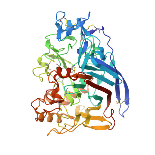
    5W11

  • PubMed Abstract: 

    Cellobiohydrolases hydrolyze cellulose, a linear polymer with glucose monomers linked exclusively by β-1,4 glycosidic linkages. The widespread hydrogen bonding network tethers individual cellulose polymers forming crystalline cellulose, which prevent the access of hydrolytic enzymes and water molecules. The most abundant enzyme secreted by Myceliophthora thermophila M77 in response to the presence of biomass is the cellobiohydrolase MtCel7A, which is composed by a GH7-catalytic domain (CD), a linker, and a CBM1-type carbohydrate-binding module. GH7 cellobiohydrolases have been studied before, and structural models have been proposed. However, currently available GH7 crystal structures only define separate catalytic domains and/or cellulose-binding modules and do not include the full-length structures that are involved in shaping the catalytic mode of operation. In this study, we determined the 3D structure of catalytic domain using X-ray crystallography and retrieved the full-length enzyme envelope via small-angle X-ray scattering (SAXS) technique. The SAXS data reveal a tadpole-like molecular shape with a rigid linker connecting the CD and CBM. Our biochemical studies show that MtCel7A has higher catalytic efficiency and thermostability as well as lower processivity when compared to the well-studied TrCel7A from Trichoderma reesei. Based on a comparison of the crystallographic structures of CDs and their molecular dynamic simulations, we demonstrate that MtCel7A has considerably higher flexibility than TrCel7A. In particular, loops that cover the active site are more flexible and undergo higher conformational fluctuations, which might account for decreased processivity and enhanced enzymatic efficiency. Our statistical coupling analysis suggests co-evolution of amino acid clusters comprising the catalytic site of MtCel7A, which correlate with the steps in the catalytic cycle of the enzyme. The atomic coordinates and structural factors of MtCel7A have been deposited in the Protein Data Bank with accession number 5W11.


  • Organizational Affiliation

    São Carlos Institute of Physics, University of São Paulo, Brazil.


Macromolecules
Find similar proteins by:  (by identity cutoff)  |  3D Structure
Entity ID: 1
MoleculeChains Sequence LengthOrganismDetailsImage
Glucanase
A, B
440Thermothelomyces thermophilusMutation(s): 0 
EC: 3.2.1.176
UniProt
Find proteins for G2Q665 (Thermothelomyces thermophilus (strain ATCC 42464 / BCRC 31852 / DSM 1799))
Explore G2Q665 
Go to UniProtKB:  G2Q665
Entity Groups  
Sequence Clusters30% Identity50% Identity70% Identity90% Identity95% Identity100% Identity
UniProt GroupG2Q665
Sequence Annotations
Expand
  • Reference Sequence
Oligosaccharides

Help

Entity ID: 2
MoleculeChains Length2D Diagram Glycosylation3D Interactions
alpha-D-mannopyranose-(1-4)-2-acetamido-2-deoxy-beta-D-glucopyranose-(1-4)-2-acetamido-2-deoxy-beta-D-glucopyranose
C, D
3N-Glycosylation
Glycosylation Resources
GlyTouCan:  G62182OO
GlyCosmos:  G62182OO
GlyGen:  G62182OO
Entity ID: 3
MoleculeChains Length2D Diagram Glycosylation3D Interactions
beta-D-glucopyranose-(1-4)-beta-D-glucopyranose-(1-4)-alpha-D-glucopyranose
E, F, G, H
3N/A
Glycosylation Resources
GlyTouCan:  G03861VK
GlyCosmos:  G03861VK
GlyGen:  G03861VK
Small Molecules
Modified Residues  1 Unique
IDChains TypeFormula2D DiagramParent
PCA
Query on PCA
A, B
L-PEPTIDE LINKINGC5 H7 N O3GLN
Experimental Data & Validation

Experimental Data

  • Method: X-RAY DIFFRACTION
  • Resolution: 2.31 Å
  • R-Value Free: 0.204 
  • R-Value Work: 0.137 
  • R-Value Observed: 0.140 
  • Space Group: P 1 21 1
Unit Cell:
Length ( Å )Angle ( ˚ )
a = 61.619α = 90
b = 93.261β = 100.46
c = 69.679γ = 90
Software Package:
Software NamePurpose
PHENIXrefinement
PDB_EXTRACTdata extraction
XDSdata reduction
Aimlessdata scaling
PHASERphasing

Structure Validation

View Full Validation Report


Ligand Structure Quality Assessment 


Entry History & Funding Information

Deposition Data


Funding OrganizationLocationGrant Number
Sao Paulo Research Foundation (FAPESP)Brazil2011/20505-4

Revision History  (Full details and data files)

  • Version 1.0: 2017-12-13
    Type: Initial release
  • Version 1.1: 2017-12-20
    Changes: Database references
  • Version 1.2: 2018-02-14
    Changes: Database references
  • Version 1.3: 2019-04-17
    Changes: Author supporting evidence, Data collection
  • Version 2.0: 2020-01-01
    Changes: Author supporting evidence, Polymer sequence
  • Version 3.0: 2020-07-29
    Type: Remediation
    Reason: Carbohydrate remediation
    Changes: Advisory, Atomic model, Data collection, Derived calculations, Non-polymer description, Structure summary
  • Version 3.1: 2023-10-04
    Changes: Data collection, Database references, Refinement description, Structure summary