


Structure of MurC from Pseudomonas aeruginosa
Horanyi, P.S., Mayclin, S.J., Seattle Structural Genomics Center for Infectious Disease (SSGCID)To be published.
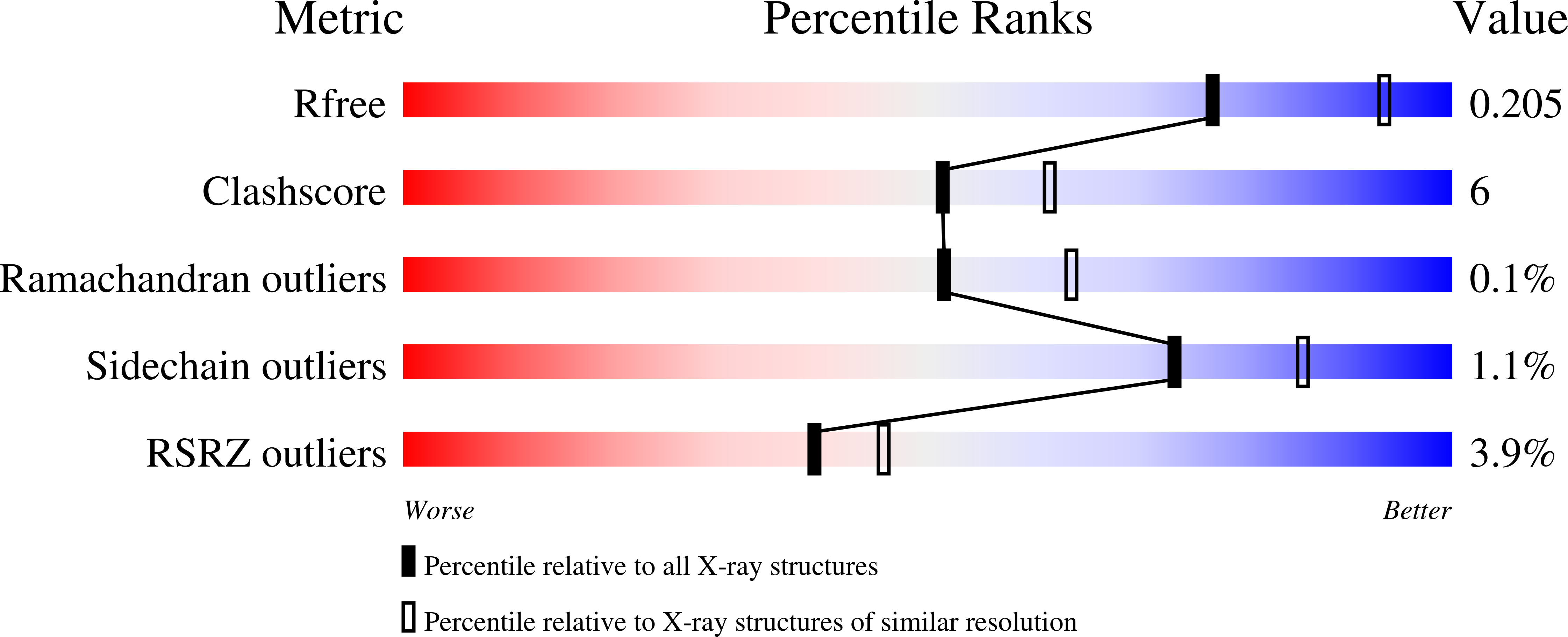
Experimental Data Snapshot
wwPDB Validation 3D Report Full Report
Entity ID: 1 | |||||
|---|---|---|---|---|---|
| Molecule | Chains | Sequence Length | Organism | Details | Image |
| UDP-N-acetylmuramate--L-alanine ligase | 315 | Pseudomonas aeruginosa PAO1 | Mutation(s): 0 Gene Names: murC, PA4411 EC: 6.3.2.8 | 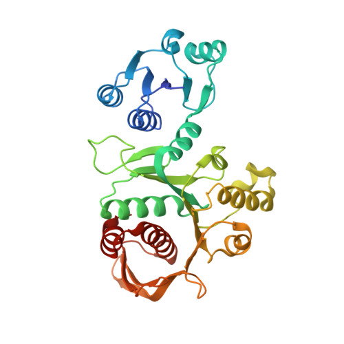 | |
UniProt | |||||
Find proteins for Q9HW02 (Pseudomonas aeruginosa (strain ATCC 15692 / DSM 22644 / CIP 104116 / JCM 14847 / LMG 12228 / 1C / PRS 101 / PAO1)) Explore Q9HW02 Go to UniProtKB: Q9HW02 | |||||
Entity Groups | |||||
| Sequence Clusters | 30% Identity50% Identity70% Identity90% Identity95% Identity100% Identity | ||||
| UniProt Group | Q9HW02 | ||||
Sequence AnnotationsExpand | |||||
| |||||
| Ligands 1 Unique | |||||
|---|---|---|---|---|---|
| ID | Chains | Name / Formula / InChI Key | 2D Diagram | 3D Interactions | |
| EDO Query on EDO | I [auth A] J [auth B] K [auth C] L [auth G] M [auth G] | 1,2-ETHANEDIOL C2 H6 O2 LYCAIKOWRPUZTN-UHFFFAOYSA-N |  | ||
| Length ( Å ) | Angle ( ˚ ) |
|---|---|
| a = 284.82 | α = 90 |
| b = 109.4 | β = 112.38 |
| c = 108.58 | γ = 90 |
| Software Name | Purpose |
|---|---|
| PHENIX | refinement |
| XDS | data reduction |
| XSCALE | data scaling |
| PHASER | phasing |
RCSB PDB (citation) is hosted by
RCSB PDB is a member of the

