5V1I

DNA polymerase beta ternary product complex with 8-oxoG:C and inserted dCTP


Experimental Data Snapshot

  • Method: X-RAY DIFFRACTION
  • Resolution: 2.04 Å
  • R-Value Free: 0.237 
  • R-Value Work: 0.184 
  • R-Value Observed: 0.188 

wwPDB Validation   3D Report Full Report


This is version 1.4 of the entry. See complete history


Literature

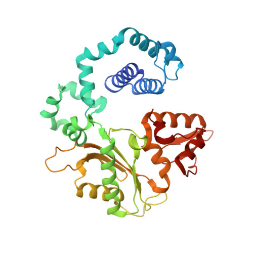
Capturing a mammalian DNA polymerase extending from an oxidized nucleotide.

Whitaker, A.M.Smith, M.R.Schaich, M.A.Freudenthal, B.D.

(2017) Nucleic Acids Res 45: 6934-6944

  • DOI: https://doi.org/10.1093/nar/gkx293
  • Primary Citation of Related Structures:  
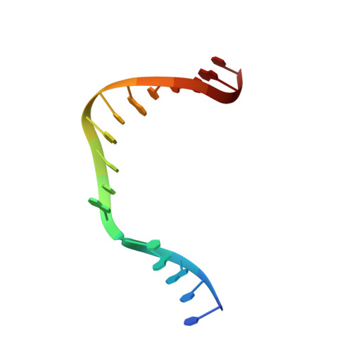
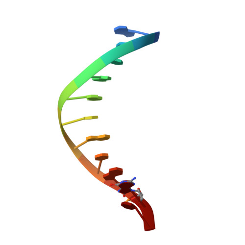
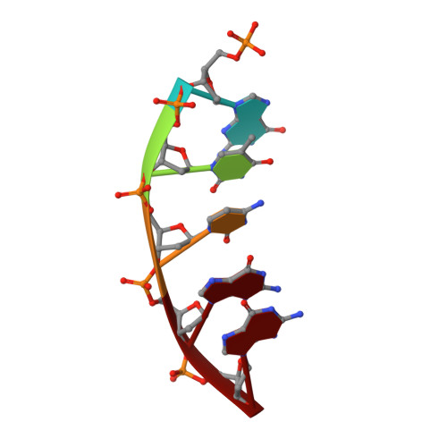
    5V1F, 5V1G, 5V1H, 5V1I, 5V1J, 5V1N, 5V1O, 5V1P, 5V1R, 5VEZ

  • PubMed Abstract: 

    The oxidized nucleotide, 8-oxo-7,8-dihydro-2΄-deoxyguanosine (8-oxoG), is one of the most abundant DNA lesions. 8-oxoG plays a major role in tumorigenesis and human disease. Biological consequences of 8-oxoG are mediated in part by its insertion into the genome, making it essential to understand how DNA polymerases handle 8-oxoG. Insertion of 8-oxoG is mutagenic when opposite adenine but not when opposite cytosine. However, either result leads to DNA damage at the primer terminus (3΄-end) during the succeeding insertion event. Extension from DNA damage at primer termini remains poorly understood. Using kinetics and time-lapse crystallography, we evaluated how a model DNA polymerase, human polymerase β, accommodates 8-oxoG at the primer terminus opposite cytosine and adenine. Notably, extension from the mutagenic base pair is favored over the non-mutagenic base pair. When 8-oxoG is at the primer terminus opposite cytosine, DNA centric changes lead to a clash between O8 of 8-oxoG and the phosphate backbone. Changes in the extension reaction resulting from the altered active site provide evidence for a stabilizing interaction between Arg254 and Asp256 that serves an important role during DNA synthesis reactions. These results provide novel insights into the impact of damage at the primer terminus on genomic stability and DNA synthesis.


  • Organizational Affiliation

    Department of Biochemistry and Molecular Biology, University of Kansas Medical Center, Kansas City, KS 66160, USA.


Macromolecules

Find similar proteins by:  (by identity cutoff)  |  3D Structure
Entity ID: 4
MoleculeChains Sequence LengthOrganismDetailsImage
DNA polymerase betaD [auth A]335Homo sapiensMutation(s): 0 
Gene Names: POLB
EC: 2.7.7.7 (PDB Primary Data), 4.2.99 (PDB Primary Data)
UniProt & NIH Common Fund Data Resources
Find proteins for P06746 (Homo sapiens)
Explore P06746 
Go to UniProtKB:  P06746
PHAROS:  P06746
GTEx:  ENSG00000070501 
Entity Groups  
Sequence Clusters30% Identity50% Identity70% Identity90% Identity95% Identity100% Identity
UniProt GroupP06746
Sequence Annotations
Expand
  • Reference Sequence

Find similar nucleic acids by:  Sequence   |   3D Structure  

Entity ID: 1
MoleculeChains LengthOrganismImage
DNA (5'-D(*CP*CP*GP*AP*CP*GP*CP*CP*GP*CP*AP*TP*CP*AP*GP*C)-3')A [auth T]16synthetic construct
Sequence Annotations
Expand
  • Reference Sequence

Find similar nucleic acids by:  Sequence   |   3D Structure  

Entity ID: 2
MoleculeChains LengthOrganismImage
DNA (5'-D(*GP*CP*TP*GP*AP*TP*GP*CP*GP*(8OG)P*C)-3')B [auth P]11synthetic construct
Sequence Annotations
Expand
  • Reference Sequence

Find similar nucleic acids by:  Sequence   |   3D Structure  

Entity ID: 3
MoleculeChains LengthOrganismImage
DNA (5'-D(P*GP*TP*CP*GP*G)-3')C [auth D]5synthetic construct
Sequence Annotations
Expand
  • Reference Sequence
Small Molecules
Ligands 4 Unique
IDChains Name / Formula / InChI Key2D Diagram3D Interactions
PPV
Query on PPV

Download Ideal Coordinates CCD File 
M [auth A]PYROPHOSPHATE
H4 O7 P2
XPPKVPWEQAFLFU-UHFFFAOYSA-N
CL
Query on CL

Download Ideal Coordinates CCD File 
H [auth A],
I [auth A],
J [auth A],
K [auth A]
CHLORIDE ION
Cl
VEXZGXHMUGYJMC-UHFFFAOYSA-M
MG
Query on MG

Download Ideal Coordinates CCD File 
E [auth P],
L [auth A]
MAGNESIUM ION
Mg
JLVVSXFLKOJNIY-UHFFFAOYSA-N
NA
Query on NA

Download Ideal Coordinates CCD File 
F [auth A],
G [auth A],
N [auth A]
SODIUM ION
Na
FKNQFGJONOIPTF-UHFFFAOYSA-N
Experimental Data & Validation

Experimental Data

  • Method: X-RAY DIFFRACTION
  • Resolution: 2.04 Å
  • R-Value Free: 0.237 
  • R-Value Work: 0.184 
  • R-Value Observed: 0.188 
  • Space Group: P 1 21 1
Unit Cell:
Length ( Å )Angle ( ˚ )
a = 50.564α = 90
b = 79.798β = 107.62
c = 55.539γ = 90
Software Package:
Software NamePurpose
PHENIXrefinement
HKL-3000data scaling

Structure Validation

View Full Validation Report


Entry History & Funding Information

Deposition Data


Funding OrganizationLocationGrant Number
National Institutes of Health/National Institute of General Medical Sciences (NIH/NIGMS)United States--

Revision History  (Full details and data files)

  • Version 1.0: 2017-05-10
    Type: Initial release
  • Version 1.1: 2017-09-13
    Changes: Database references
  • Version 1.2: 2018-04-04
    Changes: Author supporting evidence, Data collection
  • Version 1.3: 2020-01-01
    Changes: Author supporting evidence
  • Version 1.4: 2023-10-04
    Changes: Data collection, Database references, Derived calculations, Refinement description