


The quaternary structure of beta-1,3-glucan contributes to its recognition and hydrolysis by a multimodular family 81 glycoside hydrolase
Pluvinage, B., Fillo, A., Massel, P., Boraston, A.B.(2017) Structure
Experimental Data Snapshot
Starting Model: experimental
View more details
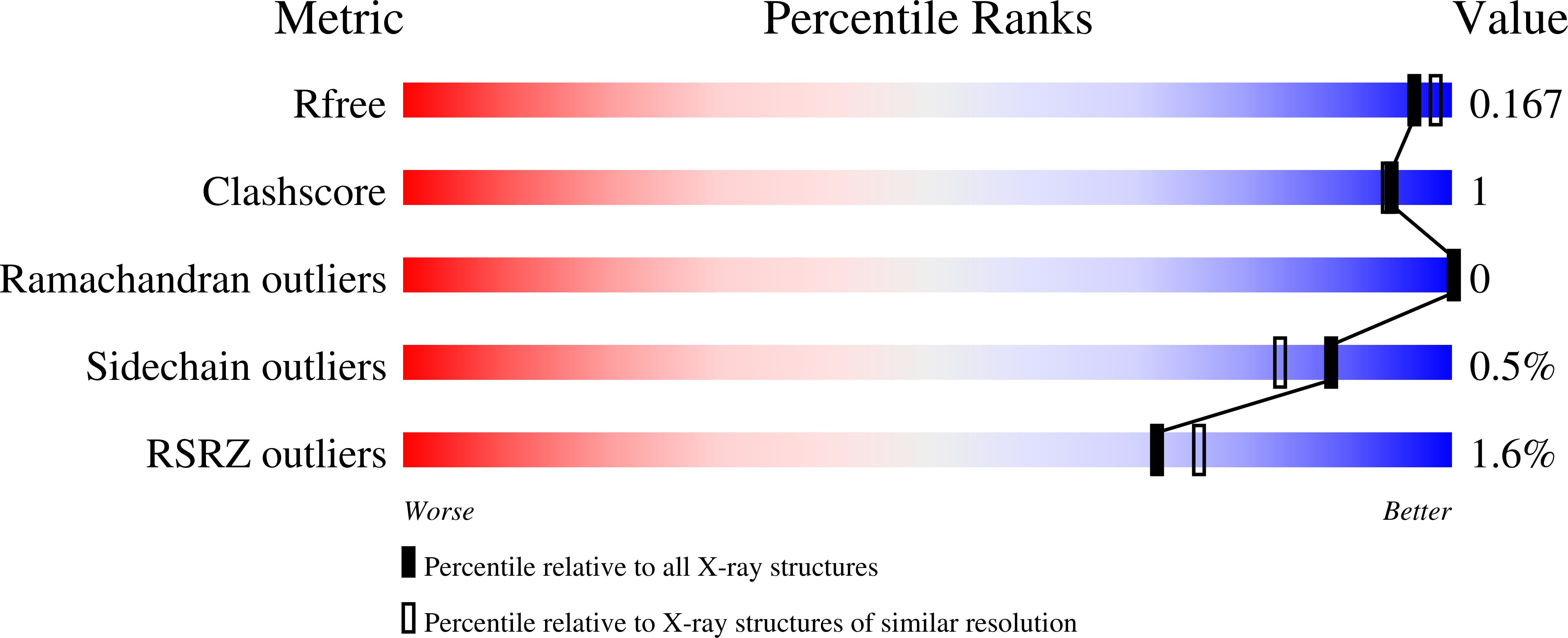
wwPDB Validation 3D Report Full Report
(2017) Structure
Entity ID: 1 | |||||
|---|---|---|---|---|---|
| Molecule | Chains | Sequence Length | Organism | Details | Image |
| BH0236 protein | 753 | Halalkalibacterium halodurans C-125 | Mutation(s): 1 Gene Names: BH0236 | 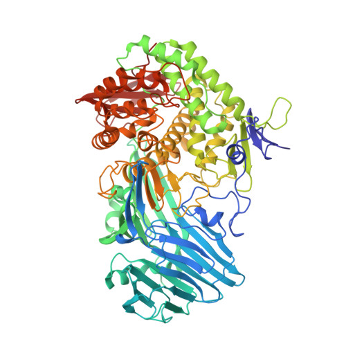 | |
UniProt | |||||
Find proteins for Q9KG76 (Halalkalibacterium halodurans (strain ATCC BAA-125 / DSM 18197 / FERM 7344 / JCM 9153 / C-125)) Explore Q9KG76 Go to UniProtKB: Q9KG76 | |||||
Entity Groups | |||||
| Sequence Clusters | 30% Identity50% Identity70% Identity90% Identity95% Identity100% Identity | ||||
| UniProt Group | Q9KG76 | ||||
Sequence AnnotationsExpand | |||||
| |||||
Entity ID: 2 | |||||
|---|---|---|---|---|---|
| Molecule | Chains | Length | 2D Diagram | Glycosylation | 3D Interactions |
| beta-D-glucopyranose-(1-3)-beta-D-glucopyranose-(1-3)-beta-D-glucopyranose | B | 3 |  | N/A | |
Glycosylation Resources | |||||
GlyTouCan: G00024MO GlyCosmos: G00024MO GlyGen: G00024MO | |||||
| Ligands 2 Unique | |||||
|---|---|---|---|---|---|
| ID | Chains | Name / Formula / InChI Key | 2D Diagram | 3D Interactions | |
| PO4 Query on PO4 | E [auth A] F [auth A] G [auth A] H [auth A] I [auth A] | PHOSPHATE ION O4 P NBIIXXVUZAFLBC-UHFFFAOYSA-K |  | ||
| EDO Query on EDO | N [auth A] O [auth A] P [auth A] Q [auth A] R [auth A] | 1,2-ETHANEDIOL C2 H6 O2 LYCAIKOWRPUZTN-UHFFFAOYSA-N |  | ||
Entity ID: 4 | |||||
|---|---|---|---|---|---|
| ID | Chains | Name | Type/Class | 2D Diagram | 3D Interactions |
| PRD_900024 Query on PRD_900024 | D | beta-laminaribiose | Oligosaccharide / Antimicrobial |  | |
| Length ( Å ) | Angle ( ˚ ) |
|---|---|
| a = 65.52 | α = 90 |
| b = 93.68 | β = 90 |
| c = 159.41 | γ = 90 |
| Software Name | Purpose |
|---|---|
| REFMAC | refinement |
| SCALA | data scaling |
| PHASER | phasing |
| PDB_EXTRACT | data extraction |
| MOSFLM | data reduction |
RCSB PDB (citation) is hosted by
RCSB PDB is a member of the


