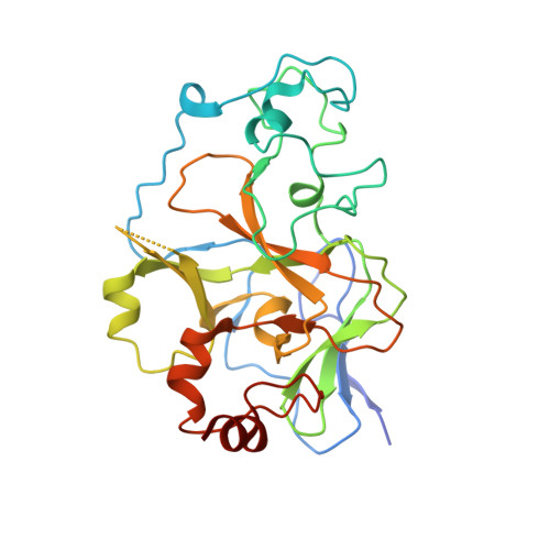
5TTF

Crystal structure of catalytic domain of G9a with MS012


Experimental Data Snapshot

  • Method: X-RAY DIFFRACTION
  • Resolution: 1.72 Å
  • R-Value Free: 0.247 
  • R-Value Work: 0.207 
  • R-Value Observed: 0.207 

wwPDB Validation   3D Report Full Report


Ligand Structure Quality Assessment 


This is version 1.2 of the entry. See complete history


Literature

Discovery of Potent and Selective Inhibitors for G9a-Like Protein (GLP) Lysine Methyltransferase.

Xiong, Y.Li, F.Babault, N.Dong, A.Zeng, H.Wu, H.Chen, X.Arrowsmith, C.H.Brown, P.J.Liu, J.Vedadi, M.Jin, J.

(2017) J Med Chem 60: 1876-1891

  • DOI: https://doi.org/10.1021/acs.jmedchem.6b01645
  • Primary Citation of Related Structures:  
    5TTF, 5TTG, 5TUY, 5TUZ

  • PubMed Abstract: 

    G9a-like protein (GLP) and G9a are highly homologous protein lysine methyltransferases (PKMTs) sharing approximately 80% sequence identity in their catalytic domains. GLP and G9a form a heterodimer complex and catalyze mono- and dimethylation of histone H3 lysine 9 and nonhistone substrates. Although they are closely related, GLP and G9a possess distinct physiological and pathophysiological functions. Thus, GLP or G9a selective small-molecule inhibitors are useful tools to dissect their distinct biological functions. We previously reported potent and selective G9a/GLP dual inhibitors including UNC0638 and UNC0642. Here we report the discovery of potent and selective GLP inhibitors including 4 (MS0124) and 18 (MS012), which are >30-fold and 140-fold selective for GLP over G9a and other methyltransferases, respectively. The cocrystal structures of GLP and G9a in the complex with either 4 or 18 displayed virtually identical binding modes and interactions, highlighting the challenges in structure-based design of selective inhibitors for either enzyme.


  • Organizational Affiliation

    Department of Pharmacological Sciences and Department of Oncological Sciences, Icahn School of Medicine at Mount Sinai , New York, New York 10029, United States.


Macromolecules
Find similar proteins by:  (by identity cutoff)  |  3D Structure
Entity ID: 1
MoleculeChains Sequence LengthOrganismDetailsImage
Histone-lysine N-methyltransferase EHMT2
A, B, C, D
283Homo sapiensMutation(s): 0 
Gene Names: EHMT2BAT8C6orf30G9AKMT1CNG36
EC: 2.1.1 (PDB Primary Data), 2.1.1.43 (PDB Primary Data)
UniProt & NIH Common Fund Data Resources
Find proteins for Q96KQ7 (Homo sapiens)
Explore Q96KQ7 
Go to UniProtKB:  Q96KQ7
PHAROS:  Q96KQ7
GTEx:  ENSG00000204371 
Entity Groups  
Sequence Clusters30% Identity50% Identity70% Identity90% Identity95% Identity100% Identity
UniProt GroupQ96KQ7
Sequence Annotations
Expand
  • Reference Sequence
Small Molecules
Ligands 5 Unique
IDChains Name / Formula / InChI Key2D Diagram3D Interactions
7KZ
Query on 7KZ

Download Ideal Coordinates CCD File 
GA [auth D],
J [auth A],
R [auth B],
Z [auth C]
N4-(1-methylpiperidin-4-yl)-N2-hexyl-6,7-dimethoxyquinazoline-2,4-diamine
C22 H35 N5 O2
ZYNUWSFRZCRKSN-UHFFFAOYSA-N
SAM
Query on SAM

Download Ideal Coordinates CCD File 
FA [auth D],
I [auth A],
Q [auth B],
Y [auth C]
S-ADENOSYLMETHIONINE
C15 H22 N6 O5 S
MEFKEPWMEQBLKI-FCKMPRQPSA-N
ZN
Query on ZN

Download Ideal Coordinates CCD File 
BA [auth D]
CA [auth D]
DA [auth D]
E [auth A]
EA [auth D]
BA [auth D],
CA [auth D],
DA [auth D],
E [auth A],
EA [auth D],
F [auth A],
G [auth A],
H [auth A],
M [auth B],
N [auth B],
O [auth B],
P [auth B],
U [auth C],
V [auth C],
W [auth C],
X [auth C]
ZINC ION
Zn
PTFCDOFLOPIGGS-UHFFFAOYSA-N
CL
Query on CL

Download Ideal Coordinates CCD File 
K [auth A]CHLORIDE ION
Cl
VEXZGXHMUGYJMC-UHFFFAOYSA-M
UNX
Query on UNX

Download Ideal Coordinates CCD File 
AA [auth C],
HA [auth D],
L [auth A],
S [auth B],
T [auth B]
UNKNOWN ATOM OR ION
X
Binding Affinity Annotations 
IDSourceBinding Affinity
7KZ BindingDB:  5TTF Kd: 610 (nM) from 1 assay(s)
IC50: 992 (nM) from 1 assay(s)
Experimental Data & Validation

Experimental Data

  • Method: X-RAY DIFFRACTION
  • Resolution: 1.72 Å
  • R-Value Free: 0.247 
  • R-Value Work: 0.207 
  • R-Value Observed: 0.207 
  • Space Group: P 1
  • Diffraction Data: https://doi.org/10.18430/M35TTF
Unit Cell:
Length ( Å )Angle ( ˚ )
a = 56.846α = 92.09
b = 67.749β = 90.05
c = 77.845γ = 91.8
Software Package:
Software NamePurpose
REFMACrefinement
SCALEPACKdata scaling
PDB_EXTRACTdata extraction
PHASERphasing
HKL-3000data reduction

Structure Validation

View Full Validation Report


Ligand Structure Quality Assessment 


Entry History 

Revision History  (Full details and data files)

  • Version 1.0: 2016-12-21
    Type: Initial release
  • Version 1.1: 2018-05-23
    Changes: Data collection, Database references
  • Version 1.2: 2023-10-04
    Changes: Data collection, Database references, Derived calculations, Refinement description