5TS5

Crystal structure of L-amino acid oxidase from Bothrops atrox


Experimental Data Snapshot

  • Method: X-RAY DIFFRACTION
  • Resolution: 2.30 Å
  • R-Value Free: 0.230 
  • R-Value Work: 0.180 
  • R-Value Observed: 0.183 

wwPDB Validation   3D Report Full Report


Ligand Structure Quality Assessment 


This is version 2.2 of the entry. See complete history


Literature

Crystal structure and molecular dynamics studies of L-amino acid oxidase from Bothrops atrox.

Feliciano, P.R.Rustiguel, J.K.Soares, R.O.Sampaio, S.V.Cristina Nonato, M.

(2017) Toxicon 128: 50-59

  • DOI: https://doi.org/10.1016/j.toxicon.2017.01.017
  • Primary Citation of Related Structures:  
    5TS5

  • PubMed Abstract: 

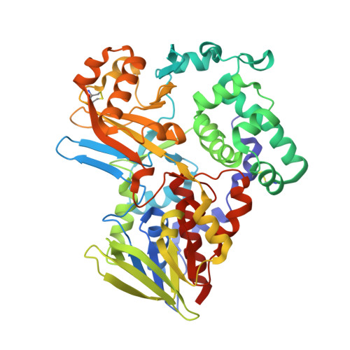
    L-amino acid oxidases (LAAOs) are dimeric flavoproteins that catalyze the deamination of L-amino acid to α-keto acid, producing ammonia and hydrogen peroxide. In this study, we report the crystal structure and molecular dynamics simulations of LAAO from the venom of Bothrops atrox (BatroxLAAO). BatroxLAAO presents several biological and pharmacological properties with promising biomedical applications. BatroxLAAO structure contains the highly conserved structural pattern of LAAOs comprising a FAD-binding domain, substrate-binding domain and helical domain, and a dimeric arrangement that can be stabilized by zinc. Also, molecular dynamics results show an asymmetric behavior, and a direct communication between FAD- and substrate-binding domains of counterpart subunits. These findings shed light on the structural role of dimerization to catalytic mechanism of SV-LAAOs.


  • Organizational Affiliation

    Laboratório de Cristalografia de Proteínas, Faculdade de Ciências Farmacêuticas de Ribeirão Preto, Universidade de São Paulo, Ribeirão Preto, SP, Brazil.


Macromolecules
Find similar proteins by:  (by identity cutoff)  |  3D Structure
Entity ID: 1
MoleculeChains Sequence LengthOrganismDetailsImage
Amine oxidase
A, B, C, D
484Bothrops atroxMutation(s): 0 
EC: 1.4.3
UniProt
Find proteins for P0CC17 (Bothrops atrox)
Explore P0CC17 
Go to UniProtKB:  P0CC17
Entity Groups  
Sequence Clusters30% Identity50% Identity70% Identity90% Identity95% Identity100% Identity
UniProt GroupP0CC17
Sequence Annotations
Expand
  • Reference Sequence
Oligosaccharides

Help

Entity ID: 2
MoleculeChains Length2D Diagram Glycosylation3D Interactions
alpha-L-fucopyranose-(1-6)-2-acetamido-2-deoxy-beta-D-glucopyranose
E, F
2N-Glycosylation
Glycosylation Resources
GlyTouCan:  G86851RC
GlyCosmos:  G86851RC
GlyGen:  G86851RC
Small Molecules
Ligands 4 Unique
IDChains Name / Formula / InChI Key2D Diagram3D Interactions
FAD
Query on FAD

Download Ideal Coordinates CCD File 
G [auth A],
M [auth B],
R [auth C],
V [auth D]
FLAVIN-ADENINE DINUCLEOTIDE
C27 H33 N9 O15 P2
VWWQXMAJTJZDQX-UYBVJOGSSA-N
NAG
Query on NAG

Download Ideal Coordinates CCD File 
J [auth A],
Q [auth B],
Y [auth D]
2-acetamido-2-deoxy-beta-D-glucopyranose
C8 H15 N O6
OVRNDRQMDRJTHS-FMDGEEDCSA-N
GOL
Query on GOL

Download Ideal Coordinates CCD File 
K [auth A]GLYCEROL
C3 H8 O3
PEDCQBHIVMGVHV-UHFFFAOYSA-N
ZN
Query on ZN

Download Ideal Coordinates CCD File 
H [auth A]
I [auth A]
L [auth A]
N [auth B]
O [auth B]
H [auth A],
I [auth A],
L [auth A],
N [auth B],
O [auth B],
P [auth B],
S [auth C],
T [auth C],
U [auth C],
W [auth D],
X [auth D]
ZINC ION
Zn
PTFCDOFLOPIGGS-UHFFFAOYSA-N
Experimental Data & Validation

Experimental Data

  • Method: X-RAY DIFFRACTION
  • Resolution: 2.30 Å
  • R-Value Free: 0.230 
  • R-Value Work: 0.180 
  • R-Value Observed: 0.183 
  • Space Group: P 1 21 1
Unit Cell:
Length ( Å )Angle ( ˚ )
a = 73.64α = 90
b = 123.92β = 96.03
c = 105.08γ = 90
Software Package:
Software NamePurpose
PHENIXrefinement
MOSFLMdata reduction
SCALAdata scaling
PHASERphasing

Structure Validation

View Full Validation Report


Ligand Structure Quality Assessment 


Entry History & Funding Information

Deposition Data


Funding OrganizationLocationGrant Number
Sao Paulo Research Foundation (FAPESP)Brazil2009/10454-3
Sao Paulo Research Foundation (FAPESP)Brazil2013/14988-8

Revision History  (Full details and data files)

  • Version 1.0: 2017-04-26
    Type: Initial release
  • Version 1.1: 2017-05-10
    Changes: Database references
  • Version 1.2: 2018-08-29
    Changes: Data collection, Database references, Structure summary
  • Version 1.3: 2019-04-17
    Changes: Author supporting evidence, Data collection
  • Version 1.4: 2020-01-01
    Changes: Author supporting evidence, Data collection
  • Version 2.0: 2020-07-29
    Type: Remediation
    Reason: Carbohydrate remediation
    Changes: Advisory, Atomic model, Data collection, Derived calculations, Structure summary
  • Version 2.1: 2023-02-01
    Changes: Advisory, Database references, Structure summary
  • Version 2.2: 2023-10-25
    Changes: Data collection, Refinement description