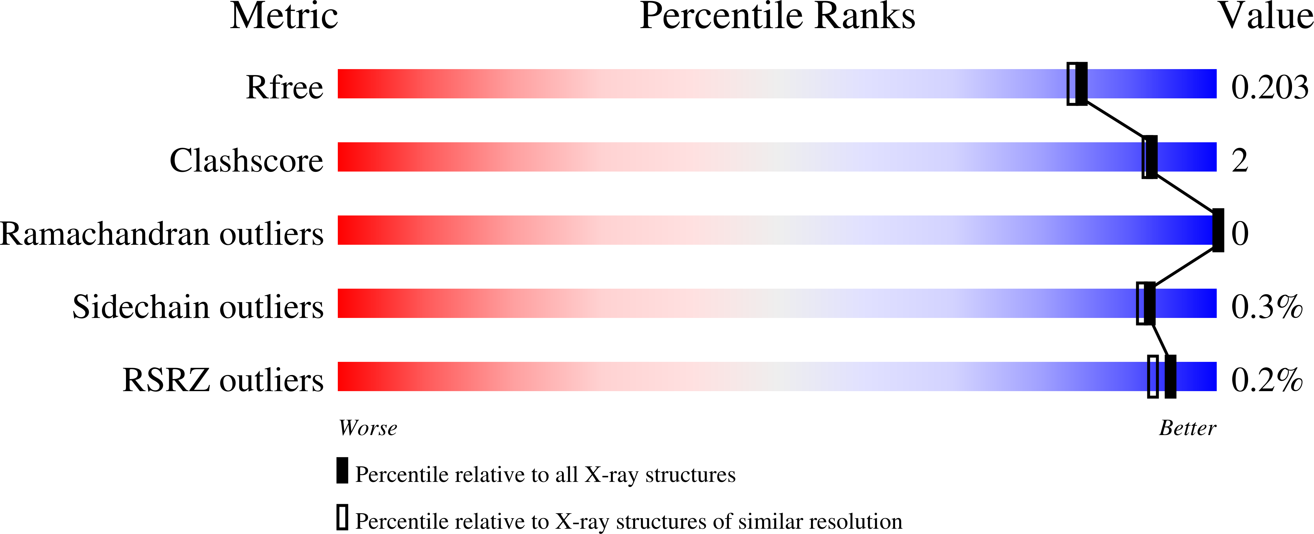
5MIH

Crystal structure of the lectin LecA from Pseudomonas aeruginosa in complex with a phenyl-epoxy-galactopyranoside


Experimental Data Snapshot

  • Method: X-RAY DIFFRACTION
  • Resolution: 1.80 Å
  • R-Value Free: 0.194 
  • R-Value Work: 0.149 
  • R-Value Observed: 0.151 

wwPDB Validation   3D Report Full Report


Ligand Structure Quality Assessment 


This is version 1.3 of the entry. See complete history


Literature

Covalent Lectin Inhibition and Application in Bacterial Biofilm Imaging.

Wagner, S.Hauck, D.Hoffmann, M.Sommer, R.Joachim, I.Muller, R.Imberty, A.Varrot, A.Titz, A.

(2017) Angew Chem Int Ed Engl 56: 16559-16564

  • DOI: https://doi.org/10.1002/anie.201709368
  • Primary Citation of Related Structures:  
    5MIH

  • PubMed Abstract: 

    Biofilm formation by pathogenic bacteria is a hallmark of chronic infections. In many cases, lectins play key roles in establishing biofilms. The pathogen Pseudomonas aeruginosa often exhibiting various drug resistances employs its lectins LecA and LecB as virulence factors and biofilm building blocks. Therefore, inhibition of the function of these proteins is thought to have potential in developing "pathoblockers" preventing biofilm formation and virulence. A covalent lectin inhibitor specific to a carbohydrate binding site is described for the first time. Its application in the LecA-specific in vitro imaging of biofilms formed by P. aeruginosa is also reported.


  • Organizational Affiliation

    Chemical Biology of Carbohydrates, Helmholtz Institute for Pharmaceutical Research Saarland (HIPS), 66123, Saarbrücken, Germany.


Macromolecules
Find similar proteins by:  (by identity cutoff)  |  3D Structure
Entity ID: 1
MoleculeChains Sequence LengthOrganismDetailsImage
PA-I galactophilic lectin
A, B, C, D
121Pseudomonas aeruginosa PAO1Mutation(s): 0 
Gene Names: lecApa1LPA2570
UniProt
Find proteins for Q05097 (Pseudomonas aeruginosa (strain ATCC 15692 / DSM 22644 / CIP 104116 / JCM 14847 / LMG 12228 / 1C / PRS 101 / PAO1))
Explore Q05097 
Go to UniProtKB:  Q05097
Entity Groups  
Sequence Clusters30% Identity50% Identity70% Identity90% Identity95% Identity100% Identity
UniProt GroupQ05097
Sequence Annotations
Expand
  • Reference Sequence
Small Molecules
Ligands 3 Unique
IDChains Name / Formula / InChI Key2D Diagram3D Interactions
7NU
Query on 7NU

Download Ideal Coordinates CCD File 
F [auth A],
H [auth B],
J [auth C],
M [auth D]
phenyl 6,7-anhydro-D-glycero-beta-D-galacto-heptopyranoside
C13 H16 O6
BGULDTQQEDQGRU-LREJFELKSA-N
ETE
Query on ETE

Download Ideal Coordinates CCD File 
K [auth C]2-{2-[2-2-(METHOXY-ETHOXY)-ETHOXY]-ETHOXY}-ETHANOL
C9 H20 O5
ZNYRFEPBTVGZDN-UHFFFAOYSA-N
CA
Query on CA

Download Ideal Coordinates CCD File 
E [auth A],
G [auth B],
I [auth C],
L [auth D]
CALCIUM ION
Ca
BHPQYMZQTOCNFJ-UHFFFAOYSA-N
Binding Affinity Annotations 
IDSourceBinding Affinity
7NU Binding MOAD:  5MIH IC50: 6.40e+4 (nM) from 1 assay(s)
Experimental Data & Validation

Experimental Data

  • Method: X-RAY DIFFRACTION
  • Resolution: 1.80 Å
  • R-Value Free: 0.194 
  • R-Value Work: 0.149 
  • R-Value Observed: 0.151 
  • Space Group: P 1 21 1
Unit Cell:
Length ( Å )Angle ( ˚ )
a = 27.343α = 90
b = 73.771β = 93.36
c = 101.454γ = 90
Software Package:
Software NamePurpose
REFMACrefinement
XDSdata reduction
Aimlessdata scaling
PHASERphasing

Structure Validation

View Full Validation Report


Ligand Structure Quality Assessment 


Entry History 

Deposition Data

Revision History  (Full details and data files)

  • Version 1.0: 2017-10-11
    Type: Initial release
  • Version 1.1: 2018-01-03
    Changes: Database references
  • Version 1.2: 2020-07-29
    Type: Remediation
    Reason: Carbohydrate remediation
    Changes: Derived calculations, Structure summary
  • Version 1.3: 2024-01-17
    Changes: Data collection, Database references, Refinement description, Structure summary