5M6E

Small Molecule inhibitors of IAP


Experimental Data Snapshot

  • Method: X-RAY DIFFRACTION
  • Resolution: 2.32 Å
  • R-Value Free: 0.239 
  • R-Value Work: 0.183 
  • R-Value Observed: 0.186 

wwPDB Validation   3D Report Full Report


Ligand Structure Quality Assessment 


This is version 2.0 of the entry. See complete history


Literature

Discovery of a Potent Nonpeptidomimetic, Small-Molecule Antagonist of Cellular Inhibitor of Apoptosis Protein 1 (cIAP1) and X-Linked Inhibitor of Apoptosis Protein (XIAP).

Tamanini, E.Buck, I.M.Chessari, G.Chiarparin, E.Day, J.E.H.Frederickson, M.Griffiths-Jones, C.M.Hearn, K.Heightman, T.D.Iqbal, A.Johnson, C.N.Lewis, E.J.Martins, V.Peakman, T.Reader, M.Rich, S.J.Ward, G.A.Williams, P.A.Wilsher, N.E.

(2017) J Med Chem 60: 4611-4625

  • DOI: https://doi.org/10.1021/acs.jmedchem.6b01877
  • Primary Citation of Related Structures:  
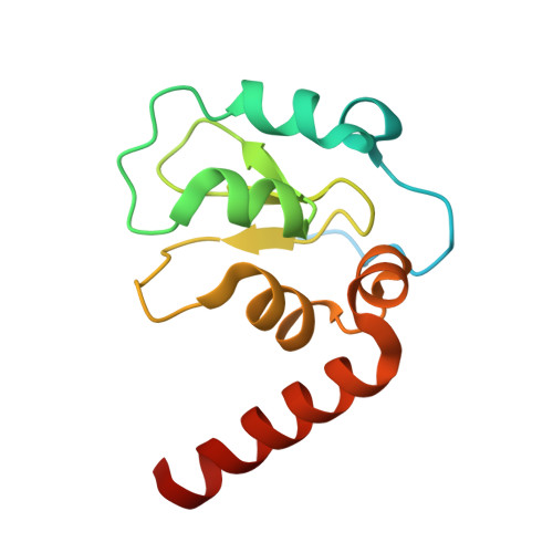
    5M6E, 5M6F, 5M6H, 5M6L, 5M6M, 5M6N

  • PubMed Abstract: 

    XIAP and cIAP1 are members of the inhibitor of apoptosis protein (IAP) family and are key regulators of anti-apoptotic and pro-survival signaling pathways. Overexpression of IAPs occurs in various cancers and has been associated with tumor progression and resistance to treatment. Structure-based drug design (SBDD) guided by structural information from X-ray crystallography, computational studies, and NMR solution conformational analysis was successfully applied to a fragment-derived lead resulting in AT-IAP, a potent, orally bioavailable, dual antagonist of XIAP and cIAP1 and a structurally novel chemical probe for IAP biology.


  • Organizational Affiliation

    Astex Pharmaceuticals , 436 Cambridge Science Park, Milton Road, Cambridge CB4 0QA, U.K.


Macromolecules
Find similar proteins by:  (by identity cutoff)  |  3D Structure
Entity ID: 1
MoleculeChains Sequence LengthOrganismDetailsImage
E3 ubiquitin-protein ligase XIAP127Homo sapiensMutation(s): 0 
Gene Names: XIAPAPI3BIRC4IAP3
EC: 6.3.2
UniProt & NIH Common Fund Data Resources
Find proteins for P98170 (Homo sapiens)
Explore P98170 
Go to UniProtKB:  P98170
PHAROS:  P98170
GTEx:  ENSG00000101966 
Entity Groups  
Sequence Clusters30% Identity50% Identity70% Identity90% Identity95% Identity100% Identity
UniProt GroupP98170
Sequence Annotations
Expand
  • Reference Sequence
Small Molecules
Ligands 4 Unique
IDChains Name / Formula / InChI Key2D Diagram3D Interactions
7HT
Query on 7HT

Download Ideal Coordinates CCD File 
E [auth A]1-[3,3-dimethyl-6-(phenylmethyl)-2~{H}-pyrrolo[3,2-c]pyridin-1-yl]-2-[(2~{R},5~{R})-5-methyl-2-[(4-methylpyrazol-1-yl)methyl]piperazin-4-ium-1-yl]ethanone
C28 H37 N6 O
CLHWAAAYYHMVJX-ZJSXRUAMSA-O
DMS
Query on DMS

Download Ideal Coordinates CCD File 
D [auth A]DIMETHYL SULFOXIDE
C2 H6 O S
IAZDPXIOMUYVGZ-UHFFFAOYSA-N
ZN
Query on ZN

Download Ideal Coordinates CCD File 
B [auth A]ZINC ION
Zn
PTFCDOFLOPIGGS-UHFFFAOYSA-N
NA
Query on NA

Download Ideal Coordinates CCD File 
C [auth A]SODIUM ION
Na
FKNQFGJONOIPTF-UHFFFAOYSA-N
Experimental Data & Validation

Experimental Data

  • Method: X-RAY DIFFRACTION
  • Resolution: 2.32 Å
  • R-Value Free: 0.239 
  • R-Value Work: 0.183 
  • R-Value Observed: 0.186 
  • Space Group: P 41 2 2
Unit Cell:
Length ( Å )Angle ( ˚ )
a = 71.525α = 90
b = 71.525β = 90
c = 105.693γ = 90
Software Package:
Software NamePurpose
REFMACrefinement
XDSdata reduction
SCALAdata scaling
REFMACphasing

Structure Validation

View Full Validation Report


Ligand Structure Quality Assessment 


Entry History 

Deposition Data

Revision History  (Full details and data files)

  • Version 1.0: 2017-05-24
    Type: Initial release
  • Version 2.0: 2017-06-28
    Changes: Database references, Non-polymer description, Structure summary