5M3Z

Crystal structure of Citrobacter freundii methionine gamma-lyase with C115H replacement in the complex with L-norleucine


Experimental Data Snapshot

  • Method: X-RAY DIFFRACTION
  • Resolution: 1.45 Å
  • R-Value Free: 0.169 
  • R-Value Work: 0.128 
  • R-Value Observed: 0.130 

wwPDB Validation   3D Report Full Report


Ligand Structure Quality Assessment 


This is version 1.2 of the entry. See complete history


Literature

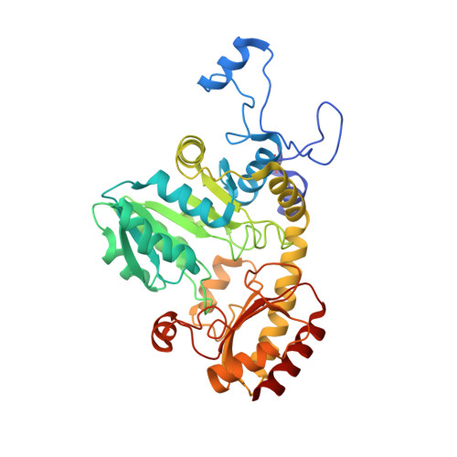
Crystal structure of mutant form Cys115His of Citrobacter freundii methionine gamma-lyase complexed with l-norleucine.

Revtovich, S.V.Morozova, E.A.Kulikova, V.V.Anufrieva, N.V.Osipova, T.I.Koval, V.S.Nikulin, A.D.Demidkina, T.V.

(2017) Biochim Biophys Acta 1865: 1123-1128

  • DOI: https://doi.org/10.1016/j.bbapap.2017.06.001
  • Primary Citation of Related Structures:  
    5M3Z

  • PubMed Abstract: 

    The mutant form of Citrobacter freundii methionine γ-lyase with the replacement of active site Cys115 for His has been found to be inactive in the γ-elimination reaction of methionine while fully active in the γ-elimination reaction of O-acetyl-l-homoserine and in the β-elimination reaction of S-alk(en)yl-substituted cysteines. In this work, the crystal structure of the mutant enzyme complexed with competitive inhibitor, l-norleucine was determined at 1.45Å resolution. At the enzyme active site the inhibitor proved to be bound both noncovalently and covalently, which corresponds to the two intermediates of the γ- and β-elimination reactions, Michaelis complex and the external aldimine. Analysis of the structure allowed us to suggest the possible reason for the inability of the mutant enzyme to catalyze the physiological reaction.


  • Organizational Affiliation

    Engelhardt Institute of Molecular Biology, Russian Academy of Sciences, Moscow, Russia.


Macromolecules
Find similar proteins by:  (by identity cutoff)  |  3D Structure
Entity ID: 1
MoleculeChains Sequence LengthOrganismDetailsImage
Methionine gamma-lyase398Citrobacter freundiiMutation(s): 1 
Gene Names: AN672_11005HMPREF3212_02785TN42_08855TO64_18625
EC: 4.4.1.11
UniProt
Find proteins for A0A0A5P8W7 (Citrobacter freundii)
Explore A0A0A5P8W7 
Go to UniProtKB:  A0A0A5P8W7
Entity Groups  
Sequence Clusters30% Identity50% Identity70% Identity90% Identity95% Identity100% Identity
UniProt GroupA0A0A5P8W7
Sequence Annotations
Expand
  • Reference Sequence
Small Molecules
Modified Residues  1 Unique
IDChains TypeFormula2D DiagramParent
CSO
Query on CSO
A
L-PEPTIDE LINKINGC3 H7 N O3 SCYS
Binding Affinity Annotations 
IDSourceBinding Affinity
NLE Binding MOAD:  5M3Z Ki: 6.00e+5 (nM) from 1 assay(s)
Experimental Data & Validation

Experimental Data

  • Method: X-RAY DIFFRACTION
  • Resolution: 1.45 Å
  • R-Value Free: 0.169 
  • R-Value Work: 0.128 
  • R-Value Observed: 0.130 
  • Space Group: I 2 2 2
Unit Cell:
Length ( Å )Angle ( ˚ )
a = 56.513α = 90
b = 123.019β = 90
c = 127.577γ = 90
Software Package:
Software NamePurpose
REFMACrefinement
XDSdata reduction
SCALAdata scaling
PHASERphasing

Structure Validation

View Full Validation Report


Ligand Structure Quality Assessment 


Entry History & Funding Information

Deposition Data


Funding OrganizationLocationGrant Number
RSFRussian Federation15-14-00009

Revision History  (Full details and data files)

  • Version 1.0: 2017-06-28
    Type: Initial release
  • Version 1.1: 2017-07-05
    Changes: Database references
  • Version 1.2: 2024-01-17
    Changes: Data collection, Database references, Refinement description