5M0W

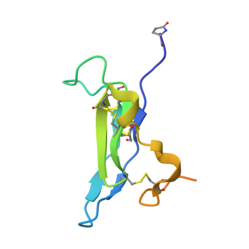
N-terminal domain of mouse Shisa 3


Experimental Data Snapshot

  • Method: X-RAY DIFFRACTION
  • Resolution: 1.39 Å
  • R-Value Free: 0.137 
  • R-Value Work: 0.115 
  • R-Value Observed: 0.116 

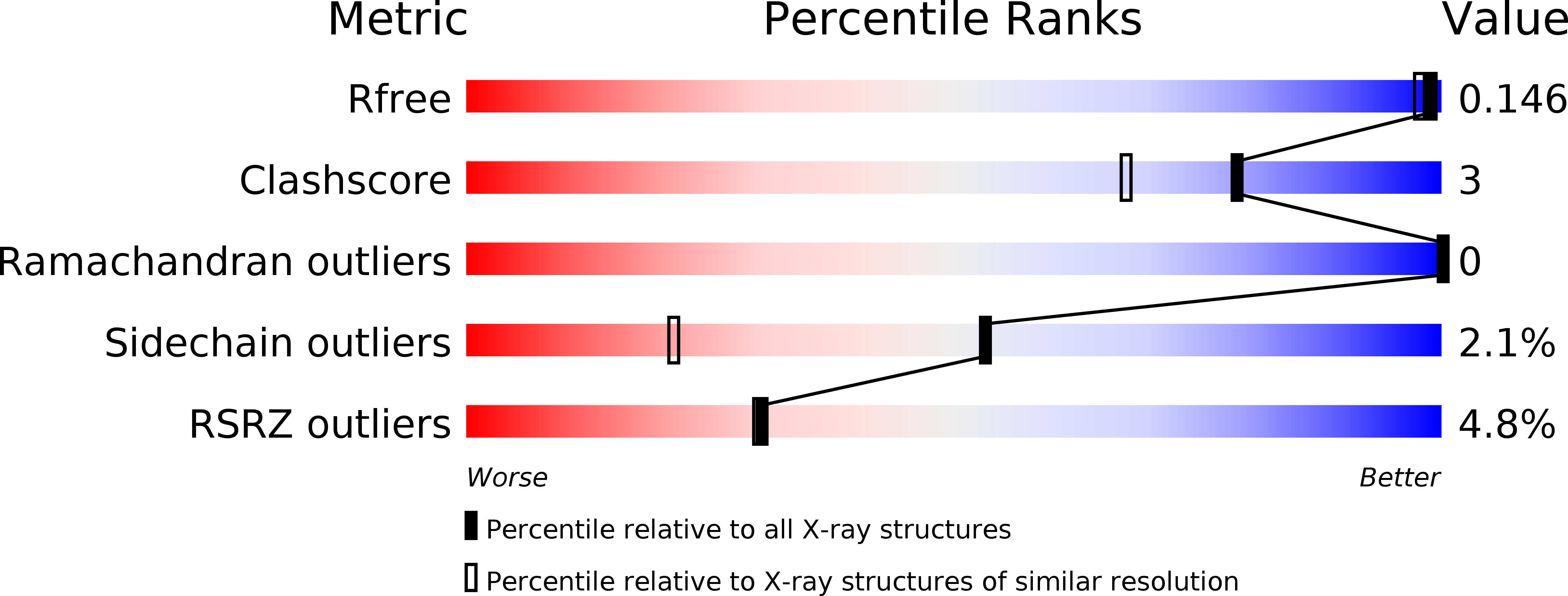
wwPDB Validation   3D Report Full Report


This is version 2.0 of the entry. See complete history


Literature

Ab initio solution of macromolecular crystal structures without direct methods.

McCoy, A.J.Oeffner, R.D.Wrobel, A.G.Ojala, J.R.Tryggvason, K.Lohkamp, B.Read, R.J.

(2017) Proc Natl Acad Sci U S A 114: 3637-3641

  • DOI: https://doi.org/10.1073/pnas.1701640114
  • Primary Citation of Related Structures:  
    5M0W

  • PubMed Abstract: 

    The majority of macromolecular crystal structures are determined using the method of molecular replacement, in which known related structures are rotated and translated to provide an initial atomic model for the new structure. A theoretical understanding of the signal-to-noise ratio in likelihood-based molecular replacement searches has been developed to account for the influence of model quality and completeness, as well as the resolution of the diffraction data. Here we show that, contrary to current belief, molecular replacement need not be restricted to the use of models comprising a substantial fraction of the unknown structure. Instead, likelihood-based methods allow a continuum of applications depending predictably on the quality of the model and the resolution of the data. Unexpectedly, our understanding of the signal-to-noise ratio in molecular replacement leads to the finding that, with data to sufficiently high resolution, fragments as small as single atoms of elements usually found in proteins can yield ab initio solutions of macromolecular structures, including some that elude traditional direct methods.


  • Organizational Affiliation

    Department of Haematology, Cambridge Institute for Medical Research, University of Cambridge, Cambridge CB2 0XY, United Kingdom.


Macromolecules
Find similar proteins by:  (by identity cutoff)  |  3D Structure
Entity ID: 1
MoleculeChains Sequence LengthOrganismDetailsImage
Protein shisa-3 homolog82Mus musculusMutation(s): 0 
Gene Names: Shisa3
UniProt
Find proteins for Q3UPR0 (Mus musculus)
Explore Q3UPR0 
Go to UniProtKB:  Q3UPR0
Entity Groups  
Sequence Clusters30% Identity50% Identity70% Identity90% Identity95% Identity100% Identity
UniProt GroupQ3UPR0
Sequence Annotations
Expand
  • Reference Sequence
Small Molecules
Ligands 5 Unique
IDChains Name / Formula / InChI Key2D Diagram3D Interactions
MES
Query on MES

Download Ideal Coordinates CCD File 
J [auth A]2-(N-MORPHOLINO)-ETHANESULFONIC ACID
C6 H13 N O4 S
SXGZJKUKBWWHRA-UHFFFAOYSA-N
PEG
Query on PEG

Download Ideal Coordinates CCD File 
K [auth A]DI(HYDROXYETHYL)ETHER
C4 H10 O3
MTHSVFCYNBDYFN-UHFFFAOYSA-N
SO4
Query on SO4

Download Ideal Coordinates CCD File 
B [auth A],
C [auth A]
SULFATE ION
O4 S
QAOWNCQODCNURD-UHFFFAOYSA-L
GOL
Query on GOL

Download Ideal Coordinates CCD File 
D [auth A],
E [auth A],
F [auth A]
GLYCEROL
C3 H8 O3
PEDCQBHIVMGVHV-UHFFFAOYSA-N
EDO
Query on EDO

Download Ideal Coordinates CCD File 
G [auth A],
H [auth A],
I [auth A]
1,2-ETHANEDIOL
C2 H6 O2
LYCAIKOWRPUZTN-UHFFFAOYSA-N
Modified Residues  1 Unique
IDChains TypeFormula2D DiagramParent
PCA
Query on PCA
A
L-PEPTIDE LINKINGC5 H7 N O3GLN
Experimental Data & Validation

Experimental Data

  • Method: X-RAY DIFFRACTION
  • Resolution: 1.39 Å
  • R-Value Free: 0.137 
  • R-Value Work: 0.115 
  • R-Value Observed: 0.116 
  • Space Group: P 43 21 2
Unit Cell:
Length ( Å )Angle ( ˚ )
a = 59.765α = 90
b = 59.765β = 90
c = 48.591γ = 90
Software Package:
Software NamePurpose
REFMACrefinement
XDSdata reduction
Aimlessdata scaling
PHASERphasing

Structure Validation

View Full Validation Report


Entry History & Funding Information

Deposition Data


Funding OrganizationLocationGrant Number
VRSweden2007-5648
Knut and Alice Wallenberg FoundationSweden--
VRSweden--
Novo Nordisk FoundationDenmark--

Revision History  (Full details and data files)

  • Version 1.0: 2017-04-05
    Type: Initial release
  • Version 1.1: 2017-04-12
    Changes: Database references
  • Version 2.0: 2020-03-11
    Changes: Derived calculations, Polymer sequence