


Crystal Structure of Human NUDT4A- Diphosphoinositol polyphosphate phosphohydrolase 2
Srikannathasan, V., Huber, K.To be published.
Experimental Data Snapshot
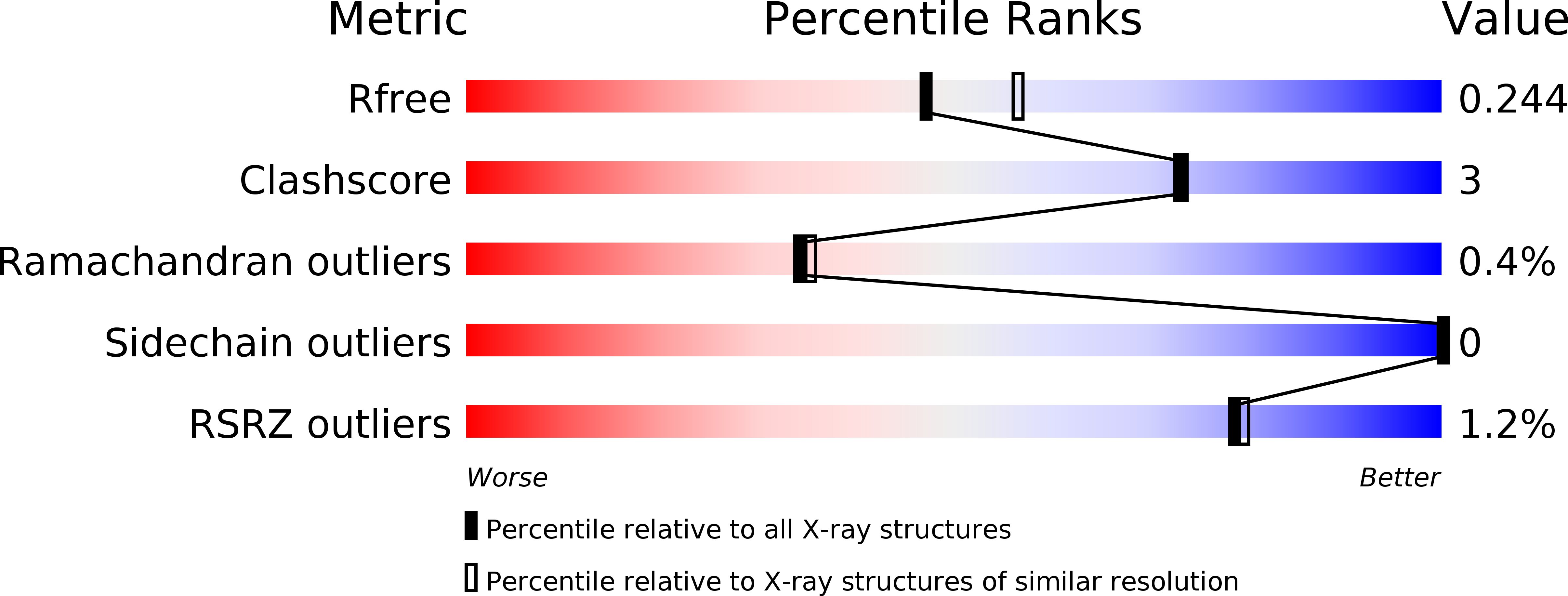
wwPDB Validation 3D Report Full Report
Entity ID: 1 | |||||
|---|---|---|---|---|---|
| Molecule | Chains | Sequence Length | Organism | Details | Image |
| Diphosphoinositol polyphosphate phosphohydrolase 2 | 180 | Homo sapiens | Mutation(s): 0 Gene Names: NUDT4, DIPP2, KIAA0487, HDCMB47P EC: 3.6.1.52 (PDB Primary Data), 3.6.1 (PDB Primary Data) |  | |
UniProt & NIH Common Fund Data Resources | |||||
Find proteins for Q9NZJ9 (Homo sapiens) Explore Q9NZJ9 Go to UniProtKB: Q9NZJ9 | |||||
PHAROS: Q9NZJ9 GTEx: ENSG00000173598 | |||||
Entity Groups | |||||
| Sequence Clusters | 30% Identity50% Identity70% Identity90% Identity95% Identity100% Identity | ||||
| UniProt Group | Q9NZJ9 | ||||
Sequence AnnotationsExpand | |||||
| |||||
| Ligands 1 Unique | |||||
|---|---|---|---|---|---|
| ID | Chains | Name / Formula / InChI Key | 2D Diagram | 3D Interactions | |
| EDO Query on EDO | C [auth A] D [auth A] E [auth A] F [auth A] G [auth A] | 1,2-ETHANEDIOL C2 H6 O2 LYCAIKOWRPUZTN-UHFFFAOYSA-N |  | ||
| Length ( Å ) | Angle ( ˚ ) |
|---|---|
| a = 135.71 | α = 90 |
| b = 41.03 | β = 122.55 |
| c = 74.32 | γ = 90 |
| Software Name | Purpose |
|---|---|
| Aimless | data scaling |
| MOLREP | phasing |
| PHENIX | refinement |
| PDB_EXTRACT | data extraction |
| Aimless | data reduction |
| MOLREP | phasing |
RCSB PDB (citation) is hosted by
RCSB PDB is a member of the

