5LOW

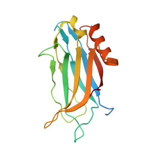
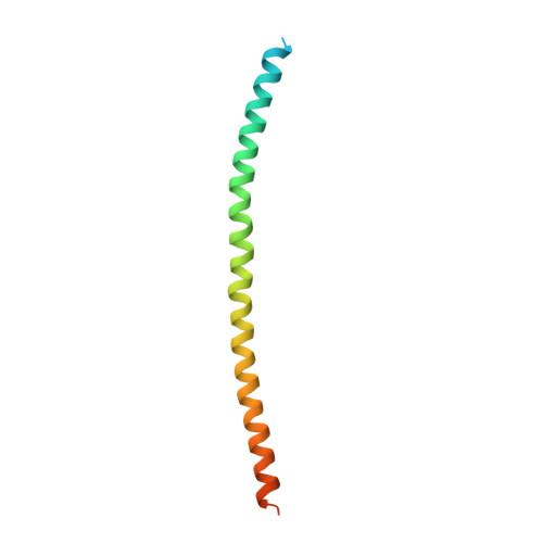
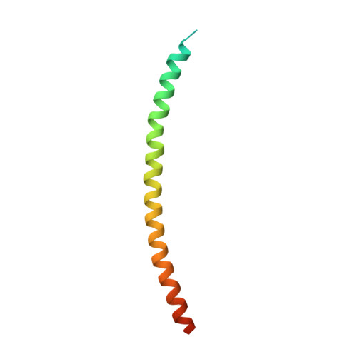
Structure of the Ca2+-bound Rabphilin 3A C2B domain SNAP25 complex (P21 space group)


Experimental Data Snapshot

  • Method: X-RAY DIFFRACTION
  • Resolution: 2.80 Å
  • R-Value Free: 0.269 
  • R-Value Work: 0.238 
  • R-Value Observed: 0.239 

wwPDB Validation   3D Report Full Report


This is version 1.2 of the entry. See complete history


Literature

Structural characterization of the Rabphilin-3A-SNAP25 interaction.

Ferrer-Orta, C.Perez-Sanchez, M.D.Coronado-Parra, T.Silva, C.Lopez-Martinez, D.Baltanas-Copado, J.Gomez-Fernandez, J.C.Corbalan-Garcia, S.Verdaguer, N.

(2017) Proc Natl Acad Sci U S A 114: E5343-E5351

  • DOI: https://doi.org/10.1073/pnas.1702542114
  • Primary Citation of Related Structures:  
    5LO8, 5LOB, 5LOW

  • PubMed Abstract: 

    Membrane fusion is essential in a myriad of eukaryotic cell biological processes, including the synaptic transmission. Rabphilin-3A is a membrane trafficking protein involved in the calcium-dependent regulation of secretory vesicle exocytosis in neurons and neuroendocrine cells, but the underlying mechanism remains poorly understood. Here, we report the crystal structures and biochemical analyses of Rabphilin-3A C2B-SNAP25 and C2B-phosphatidylinositol 4,5-bisphosphate (PIP 2 ) complexes, revealing how Rabphilin-3A C2 domains operate in cooperation with PIP 2 /Ca 2+ and SNAP25 to bind the plasma membrane, adopting a conformation compatible to interact with the complete SNARE complex. Comparisons with the synaptotagmin1-SNARE show that both proteins contact the same SNAP25 surface, but Rabphilin-3A uses a unique structural element. Data obtained here suggest a model to explain the Ca 2+ -dependent fusion process by membrane bending with a myriad of variations depending on the properties of the C2 domain-bearing protein, shedding light to understand the fine-tuning control of the different vesicle fusion events.


  • Organizational Affiliation

    Structural Biology Unit, Institut de Biologia Molecular de Barcelona, Consejo Superior de Investigaciones Cientificas, 08028 Barcelona, Spain; cfocri@ibmb.csic.es senena@um.es nvmcri@ibmb.csic.es.


Macromolecules
Find similar proteins by:  (by identity cutoff)  |  3D Structure
Entity ID: 1
MoleculeChains Sequence LengthOrganismDetailsImage
Rabphilin-3A
A, B, C, H, I
A, B, C, H, I, J
162Rattus norvegicusMutation(s): 0 
Gene Names: Rph3a
UniProt
Find proteins for P47709 (Rattus norvegicus)
Explore P47709 
Go to UniProtKB:  P47709
Entity Groups  
Sequence Clusters30% Identity50% Identity70% Identity90% Identity95% Identity100% Identity
UniProt GroupP47709
Sequence Annotations
Expand
  • Reference Sequence
Find similar proteins by:  (by identity cutoff)  |  3D Structure
Entity ID: 2
MoleculeChains Sequence LengthOrganismDetailsImage
Synaptosomal-associated protein 25
D, F, K, M
95Rattus norvegicusMutation(s): 0 
Gene Names: Snap25Snap
UniProt
Find proteins for P60881 (Rattus norvegicus)
Explore P60881 
Go to UniProtKB:  P60881
Entity Groups  
Sequence Clusters30% Identity50% Identity70% Identity90% Identity95% Identity100% Identity
UniProt GroupP60881
Sequence Annotations
Expand
  • Reference Sequence
Find similar proteins by:  (by identity cutoff)  |  3D Structure
Entity ID: 3
MoleculeChains Sequence LengthOrganismDetailsImage
Synaptosomal-associated protein 25
E, G, L, N
82Rattus norvegicusMutation(s): 0 
Gene Names: Snap25Snap
UniProt
Find proteins for P60881 (Rattus norvegicus)
Explore P60881 
Go to UniProtKB:  P60881
Entity Groups  
Sequence Clusters30% Identity50% Identity70% Identity90% Identity95% Identity100% Identity
UniProt GroupP60881
Sequence Annotations
Expand
  • Reference Sequence
Small Molecules
Ligands 2 Unique
IDChains Name / Formula / InChI Key2D Diagram3D Interactions
GOL
Query on GOL

Download Ideal Coordinates CCD File 
DA [auth K]
EA [auth M]
FA [auth M]
GA [auth N]
O [auth A]
DA [auth K],
EA [auth M],
FA [auth M],
GA [auth N],
O [auth A],
R [auth B],
U [auth C]
GLYCEROL
C3 H8 O3
PEDCQBHIVMGVHV-UHFFFAOYSA-N
CA
Query on CA

Download Ideal Coordinates CCD File 
AA [auth I]
BA [auth J]
CA [auth J]
P [auth A]
Q [auth A]
AA [auth I],
BA [auth J],
CA [auth J],
P [auth A],
Q [auth A],
S [auth B],
T [auth B],
V [auth C],
W [auth C],
X [auth H],
Y [auth H],
Z [auth I]
CALCIUM ION
Ca
BHPQYMZQTOCNFJ-UHFFFAOYSA-N
Experimental Data & Validation

Experimental Data

  • Method: X-RAY DIFFRACTION
  • Resolution: 2.80 Å
  • R-Value Free: 0.269 
  • R-Value Work: 0.238 
  • R-Value Observed: 0.239 
  • Space Group: P 1 21 1
Unit Cell:
Length ( Å )Angle ( ˚ )
a = 60.08α = 90
b = 239.496β = 114.92
c = 71.487γ = 90
Software Package:
Software NamePurpose
REFMACrefinement
SCALAdata scaling
PHASERphasing
XDSdata reduction

Structure Validation

View Full Validation Report


Entry History 

Deposition Data

Revision History  (Full details and data files)

  • Version 1.0: 2017-06-21
    Type: Initial release
  • Version 1.1: 2017-12-06
    Changes: Database references
  • Version 1.2: 2024-01-10
    Changes: Data collection, Database references, Derived calculations, Refinement description