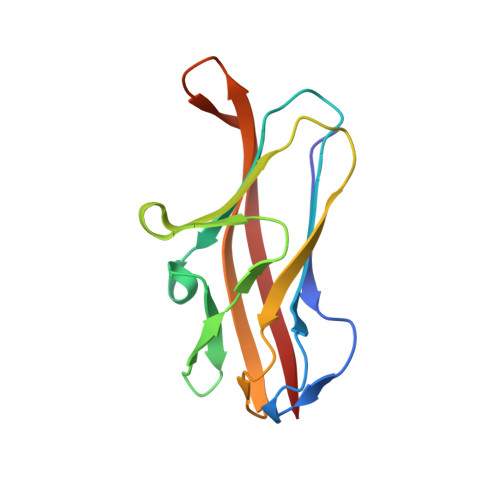
Structural basis for Myf and Psa fimbriae-mediated tropism of pathogenic strains of Yersinia for host tissues.
Pakharukova, N., Roy, S., Tuittila, M., Rahman, M.M., Paavilainen, S., Ingars, A.K., Skaldin, M., Lamminmaki, U., Hard, T., Teneberg, S., Zavialov, A.V.(2016) Mol Microbiol 102: 593-610
- PubMed: 27507539
- DOI: https://doi.org/10.1111/mmi.13481
- Primary Citation of Related Structures:
5LN4, 5LN8, 5LND, 5LO7 - PubMed Abstract:
Three pathogenic species of the genus Yersinia assemble adhesive fimbriae via the FGL-chaperone/usher pathway. Closely related Y. pestis and Y. pseudotuberculosis elaborate the pH6 antigen (Psa), which mediates bacterial attachment to alveolar cells of the lung. Y. enterocolitica, instead, assembles the homologous fimbriae Myf of unknown function. Here, we discovered that Myf, like Psa, specifically recognizes β1-3- or β1-4-linked galactose in glycosphingolipids, but completely lacks affinity for phosphatidylcholine, the main receptor for Psa in alveolar cells. The crystal structure of a subunit of Psa (PsaA) complexed with choline together with mutagenesis experiments revealed that PsaA has four phosphatidylcholine binding pockets that enable super-high-avidity binding of Psa-fibres to cell membranes. The pockets are arranged as six tyrosine residues, which are all missing in the MyfA subunit of Myf. Conversely, the crystal structure of the MyfA-galactose complex revealed that the galactose-binding site is more extended in MyfA, enabling tighter binding to lactosyl moieties. Our results suggest that during evolution, Psa has acquired a tyrosine-rich surface that enables it to bind to phosphatidylcholine and mediate adhesion of Y. pestis/pseudotuberculosis to alveolar cells, whereas Myf has specialized as a carbohydrate-binding adhesin, facilitating the attachment of Y. enterocolitica to intestinal cells.
Organizational Affiliation:
Department of Chemistry, University of Turku, Turku, Joint Biotechnology Laboratory, Arcanum, Vatselankatu 2, Turku, 20500, Finland.