5LFK

Crystal structure of CpxAHDC (hemiphosphorylated form)


Experimental Data Snapshot

  • Method: X-RAY DIFFRACTION
  • Resolution: 3.09 Å
  • R-Value Free: 0.220 
  • R-Value Work: 0.195 
  • R-Value Observed: 0.196 

wwPDB Validation   3D Report Full Report


Ligand Structure Quality Assessment 


This is version 1.2 of the entry. See complete history


Literature

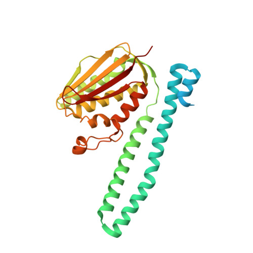
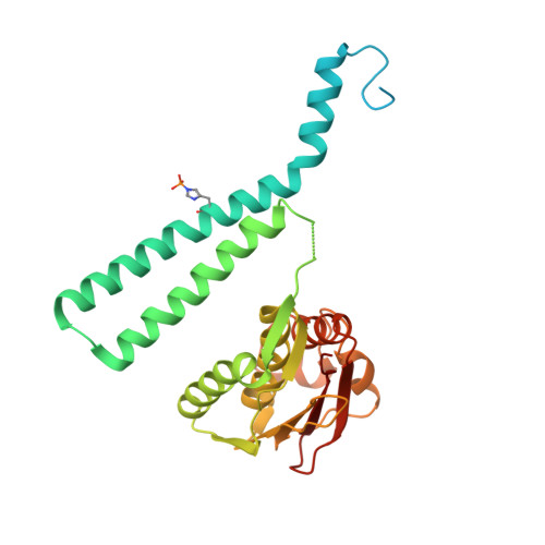
Structural Coupling between Autokinase and Phosphotransferase Reactions in a Bacterial Histidine Kinase.

Mechaly, A.E.Soto Diaz, S.Sassoon, N.Buschiazzo, A.Betton, J.M.Alzari, P.M.

(2017) Structure 25: 939-944.e3

  • DOI: https://doi.org/10.1016/j.str.2017.04.011
  • Primary Citation of Related Structures:  
    4UHJ, 4UHK, 5LFK

  • PubMed Abstract: 

    Bacterial two-component systems consist of a sensor histidine kinase (HK) and a response regulator (RR). HKs are homodimers that catalyze the autophosphorylation of a histidine residue and the subsequent phosphoryl transfer to its RR partner, triggering an adaptive response. How the HK autokinase and phosphotransferase activities are coordinated remains unclear. Here, we report X-ray structures of the prototypical HK CpxA trapped as a hemi-phosphorylated dimer, and of the receiver domain from the RR partner, CpxR. Our results reveal that the two catalytic reactions can occur simultaneously, one in each protomer of the asymmetric CpxA dimer. Furthermore, the increase of autokinase activity in the presence of phosphotransfer-impaired CpxR put forward the idea of an allosteric switching mechanism, according to which CpxR binding to one CpxA protomer triggers autophosphorylation in the second protomer. The ensuing dynamical model provides a mechanistic explanation of how HKs can efficiently orchestrate two catalytic reactions involving large-scale protein motions.


  • Organizational Affiliation

    Institut Pasteur, Unité de Microbiologie Structurale, CNRS UMR 3528 & Université Paris Diderot, Sorbonne Paris Cité, 25 rue du Dr. Roux, 75724, Paris Cedex 15, France. Electronic address: aemechaly@pasteur.edu.uy.


Macromolecules
Find similar proteins by:  (by identity cutoff)  |  3D Structure
Entity ID: 1
MoleculeChains Sequence LengthOrganismDetailsImage
Sensor histidine kinase CpxA298Escherichia coliMutation(s): 1 
Gene Names: cpxAecfBeupssdb3911JW3882
EC: 2.7.13.3
UniProt
Find proteins for P0AE82 (Escherichia coli (strain K12))
Explore P0AE82 
Go to UniProtKB:  P0AE82
Entity Groups  
Sequence Clusters30% Identity50% Identity70% Identity90% Identity95% Identity100% Identity
UniProt GroupP0AE82
Sequence Annotations
Expand
  • Reference Sequence
Find similar proteins by:  (by identity cutoff)  |  3D Structure
Entity ID: 2
MoleculeChains Sequence LengthOrganismDetailsImage
Sensor histidine kinase CpxA298Escherichia coliMutation(s): 1 
Gene Names: cpxAecfBeupssdb3911JW3882
EC: 2.7.13.3
UniProt
Find proteins for P0AE82 (Escherichia coli (strain K12))
Explore P0AE82 
Go to UniProtKB:  P0AE82
Entity Groups  
Sequence Clusters30% Identity50% Identity70% Identity90% Identity95% Identity100% Identity
UniProt GroupP0AE82
Sequence Annotations
Expand
  • Reference Sequence
Small Molecules
Ligands 3 Unique
IDChains Name / Formula / InChI Key2D Diagram3D Interactions
ATP
Query on ATP

Download Ideal Coordinates CCD File 
E [auth A],
I [auth B]
ADENOSINE-5'-TRIPHOSPHATE
C10 H16 N5 O13 P3
ZKHQWZAMYRWXGA-KQYNXXCUSA-N
SO4
Query on SO4

Download Ideal Coordinates CCD File 
C [auth A]
D [auth A]
G [auth A]
H [auth A]
K [auth B]
C [auth A],
D [auth A],
G [auth A],
H [auth A],
K [auth B],
L [auth B]
SULFATE ION
O4 S
QAOWNCQODCNURD-UHFFFAOYSA-L
MG
Query on MG

Download Ideal Coordinates CCD File 
F [auth A],
J [auth B]
MAGNESIUM ION
Mg
JLVVSXFLKOJNIY-UHFFFAOYSA-N
Modified Residues  1 Unique
IDChains TypeFormula2D DiagramParent
NEP
Query on NEP
B
L-PEPTIDE LINKINGC6 H10 N3 O5 PHIS
Experimental Data & Validation

Experimental Data

  • Method: X-RAY DIFFRACTION
  • Resolution: 3.09 Å
  • R-Value Free: 0.220 
  • R-Value Work: 0.195 
  • R-Value Observed: 0.196 
  • Space Group: P 61 2 2
Unit Cell:
Length ( Å )Angle ( ˚ )
a = 145.734α = 90
b = 145.734β = 90
c = 225.191γ = 120
Software Package:
Software NamePurpose
PHENIXrefinement
XDSdata reduction
Aimlessdata scaling
PHASERphasing

Structure Validation

View Full Validation Report


Ligand Structure Quality Assessment 


Entry History 

Deposition Data

Revision History  (Full details and data files)

  • Version 1.0: 2017-06-07
    Type: Initial release
  • Version 1.1: 2017-06-14
    Changes: Database references
  • Version 1.2: 2024-01-10
    Changes: Data collection, Database references, Derived calculations, Experimental preparation, Refinement description