5L15

The crystal structure of neuraminidase in complex with oseltamivir from A/Shanghai/2/2013 (H7N9) influenza virus


Experimental Data Snapshot

  • Method: X-RAY DIFFRACTION
  • Resolution: 2.40 Å
  • R-Value Free: 0.195 
  • R-Value Work: 0.167 
  • R-Value Observed: 0.169 

wwPDB Validation   3D Report Full Report


Ligand Structure Quality Assessment 


This is version 2.1 of the entry. See complete history


Literature

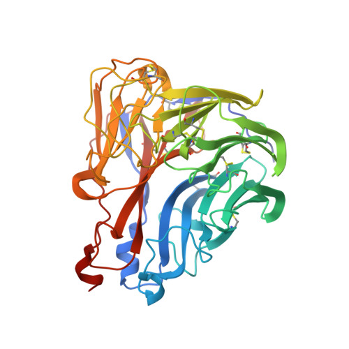
Drug Susceptibility Evaluation of an Influenza A(H7N9) Virus by Analyzing Recombinant Neuraminidase Proteins.

Gubareva, L.V.Sleeman, K.Guo, Z.Yang, H.Hodges, E.Davis, C.T.Baranovich, T.Stevens, J.

(2017) J Infect Dis 216: S566-S574

  • DOI: https://doi.org/10.1093/infdis/jiw625
  • Primary Citation of Related Structures:  
    5L14, 5L15, 5L17, 5L18

  • PubMed Abstract: 

    Neuraminidase (NA) inhibitors are the recommended antiviral medications for influenza treatment. However, their therapeutic efficacy can be compromised by NA changes that emerge naturally and/or following antiviral treatment. Knowledge of which molecular changes confer drug resistance of influenza A(H7N9) viruses (group 2NA) remains sparse.


  • Organizational Affiliation

    Influenza Division, National Center for Immunization and Respiratory Diseases, Centers for Disease Control and Prevention.


Macromolecules
Find similar proteins by:  (by identity cutoff)  |  3D Structure
Entity ID: 1
MoleculeChains Sequence LengthOrganismDetailsImage
Neuraminidase397Influenza A virus (A/Shanghai/02/2013(H7N9))Mutation(s): 0 
Gene Names: NA
EC: 3.2.1.18
UniProt
Find proteins for R4NFR6 (Influenza A virus)
Explore R4NFR6 
Go to UniProtKB:  R4NFR6
Entity Groups  
Sequence Clusters30% Identity50% Identity70% Identity90% Identity95% Identity100% Identity
UniProt GroupR4NFR6
Sequence Annotations
Expand
  • Reference Sequence
Oligosaccharides

Help

Entity ID: 2
MoleculeChains Length2D Diagram Glycosylation3D Interactions
alpha-D-mannopyranose-(1-2)-alpha-D-mannopyranose-(1-2)-alpha-D-mannopyranose-(1-3)-[alpha-D-mannopyranose-(1-3)-[alpha-D-mannopyranose-(1-6)]alpha-D-mannopyranose-(1-6)]beta-D-mannopyranose-(1-4)-2-acetamido-2-deoxy-beta-D-glucopyranose-(1-4)-2-acetamido-2-deoxy-beta-D-glucopyranose
B
9N-Glycosylation
Glycosylation Resources
GlyTouCan:  G68668TB
GlyCosmos:  G68668TB
GlyGen:  G68668TB
Small Molecules
Ligands 3 Unique
IDChains Name / Formula / InChI Key2D Diagram3D Interactions
G39
Query on G39

Download Ideal Coordinates CCD File 
E [auth A](3R,4R,5S)-4-(acetylamino)-5-amino-3-(pentan-3-yloxy)cyclohex-1-ene-1-carboxylic acid
C14 H24 N2 O4
NENPYTRHICXVCS-YNEHKIRRSA-N
NAG
Query on NAG

Download Ideal Coordinates CCD File 
C [auth A]2-acetamido-2-deoxy-beta-D-glucopyranose
C8 H15 N O6
OVRNDRQMDRJTHS-FMDGEEDCSA-N
CA
Query on CA

Download Ideal Coordinates CCD File 
D [auth A]CALCIUM ION
Ca
BHPQYMZQTOCNFJ-UHFFFAOYSA-N
Experimental Data & Validation

Experimental Data

  • Method: X-RAY DIFFRACTION
  • Resolution: 2.40 Å
  • R-Value Free: 0.195 
  • R-Value Work: 0.167 
  • R-Value Observed: 0.169 
  • Space Group: I 4 3 2
Unit Cell:
Length ( Å )Angle ( ˚ )
a = 182.436α = 90
b = 182.436β = 90
c = 182.436γ = 90
Software Package:
Software NamePurpose
PHENIXrefinement
HKL-2000data reduction
HKL-2000data scaling
PHASERphasing

Structure Validation

View Full Validation Report


Ligand Structure Quality Assessment 


Entry History 

Deposition Data

Revision History  (Full details and data files)

  • Version 1.0: 2017-08-09
    Type: Initial release
  • Version 1.1: 2017-10-04
    Changes: Database references
  • Version 2.0: 2020-07-29
    Type: Remediation
    Reason: Carbohydrate remediation
    Changes: Atomic model, Data collection, Derived calculations, Structure summary
  • Version 2.1: 2023-10-04
    Changes: Data collection, Database references, Derived calculations, Refinement description, Structure summary