5KWY

Structure of human NPC1 middle lumenal domain bound to NPC2


Experimental Data Snapshot

  • Method: X-RAY DIFFRACTION
  • Resolution: 2.40 Å
  • R-Value Free: 0.249 
  • R-Value Work: 0.194 
  • R-Value Observed: 0.197 

wwPDB Validation   3D Report Full Report


Ligand Structure Quality Assessment 


This is version 1.4 of the entry. See complete history


Literature

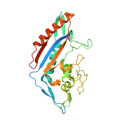
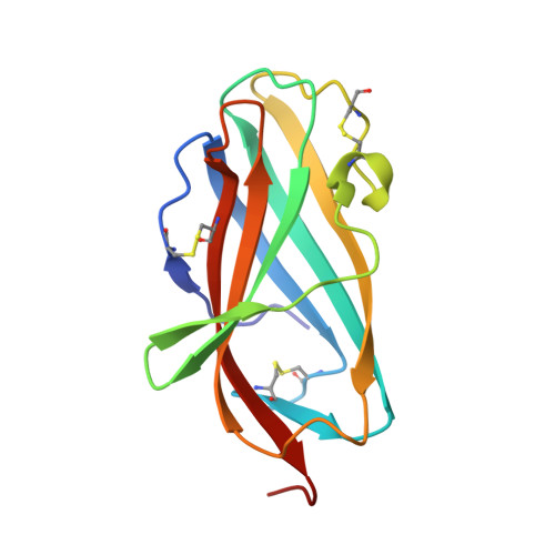
Clues to the mechanism of cholesterol transfer from the structure of NPC1 middle lumenal domain bound to NPC2.

Li, X.Saha, P.Li, J.Blobel, G.Pfeffer, S.R.

(2016) Proc Natl Acad Sci U S A 113: 10079-10084

  • DOI: https://doi.org/10.1073/pnas.1611956113
  • Primary Citation of Related Structures:  
    5KWY

  • PubMed Abstract: 

    Export of LDL-derived cholesterol from lysosomes requires the cooperation of the integral membrane protein Niemann-Pick C1 (NPC1) and a soluble protein, Niemann-Pick C2 (NPC2). Mutations in the genes encoding these proteins lead to Niemann-Pick disease type C (NPC). NPC2 binds to NPC1's second (middle), lumenally oriented domain (MLD) and transfers cholesterol to NPC1's N-terminal domain (NTD). Here, we report the 2.4-Å resolution crystal structure of a complex of human NPC1-MLD and NPC2 bearing bound cholesterol-3-O-sulfate. NPC1-MLD uses two protruding loops to bind NPC2, analogous to its interaction with the primed Ebola virus glycoprotein. Docking of the NPC1-NPC2 complex onto the full-length NPC1 structure reveals a direct cholesterol transfer tunnel between NPC2 and NTD cholesterol binding pockets, supporting the "hydrophobic hand-off" cholesterol transfer model.


  • Organizational Affiliation

    Laboratory of Cell Biology, Howard Hughes Medical Institute, The Rockefeller University, New York, NY 10065; xli05@rockefeller.edu blobel@rockefeller.edu pfeffer@stanford.edu.


Macromolecules
Find similar proteins by:  (by identity cutoff)  |  3D Structure
Entity ID: 1
MoleculeChains Sequence LengthOrganismDetailsImage
Niemann-Pick C1 protein
A, B
247Homo sapiensMutation(s): 0 
Gene Names: NPC1
UniProt & NIH Common Fund Data Resources
Find proteins for O15118 (Homo sapiens)
Explore O15118 
Go to UniProtKB:  O15118
PHAROS:  O15118
GTEx:  ENSG00000141458 
Entity Groups  
Sequence Clusters30% Identity50% Identity70% Identity90% Identity95% Identity100% Identity
UniProt GroupO15118
Sequence Annotations
Expand
  • Reference Sequence
Find similar proteins by:  (by identity cutoff)  |  3D Structure
Entity ID: 2
MoleculeChains Sequence LengthOrganismDetailsImage
Epididymal secretory protein E1
C, D
133Homo sapiensMutation(s): 0 
Gene Names: NPC2HE1
UniProt & NIH Common Fund Data Resources
Find proteins for P61916 (Homo sapiens)
Explore P61916 
Go to UniProtKB:  P61916
PHAROS:  P61916
GTEx:  ENSG00000119655 
Entity Groups  
Sequence Clusters30% Identity50% Identity70% Identity90% Identity95% Identity100% Identity
UniProt GroupP61916
Sequence Annotations
Expand
  • Reference Sequence
Experimental Data & Validation

Experimental Data

  • Method: X-RAY DIFFRACTION
  • Resolution: 2.40 Å
  • R-Value Free: 0.249 
  • R-Value Work: 0.194 
  • R-Value Observed: 0.197 
  • Space Group: C 2 2 21
Unit Cell:
Length ( Å )Angle ( ˚ )
a = 98.956α = 90
b = 109.686β = 90
c = 154.56γ = 90
Software Package:
Software NamePurpose
PHENIXrefinement
HKL-2000data reduction
HKL-2000data scaling
PHENIXphasing

Structure Validation

View Full Validation Report


Ligand Structure Quality Assessment 


Entry History 

Deposition Data

  • Released Date: 2016-08-24 
  • Deposition Author(s): Li, X.

Revision History  (Full details and data files)

  • Version 1.0: 2016-08-24
    Type: Initial release
  • Version 1.1: 2016-09-07
    Changes: Database references
  • Version 1.2: 2016-09-14
    Changes: Database references
  • Version 1.3: 2020-07-29
    Type: Remediation
    Reason: Carbohydrate remediation
    Changes: Data collection, Database references, Derived calculations, Structure summary
  • Version 1.4: 2023-10-04
    Changes: Data collection, Database references, Refinement description, Structure summary