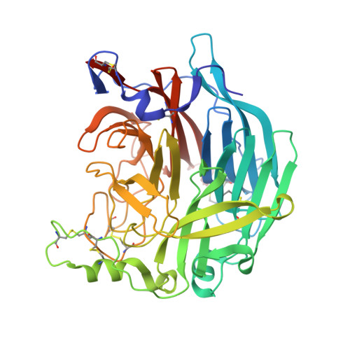
5KV8

Crystal structure of a hPIV haemagglutinin-neuraminidase-inhibitor complex


Experimental Data Snapshot

  • Method: X-RAY DIFFRACTION
  • Resolution: 1.95 Å
  • R-Value Free: 0.200 
  • R-Value Work: 0.161 
  • R-Value Observed: 0.163 

wwPDB Validation   3D Report Full Report


Ligand Structure Quality Assessment 


This is version 3.1 of the entry. See complete history


Literature

The impact of the butterfly effect on human parainfluenza virus haemagglutinin-neuraminidase inhibitor design.

Dirr, L.El-Deeb, I.M.Chavas, L.M.G.Guillon, P.Itzstein, M.V.

(2017) Sci Rep 7: 4507-4507

  • DOI: https://doi.org/10.1038/s41598-017-04656-y
  • Primary Citation of Related Structures:  
    5KV8, 5KV9

  • PubMed Abstract: 

    Human parainfluenza viruses represent a leading cause of lower respiratory tract disease in children, with currently no available approved drug or vaccine. The viral surface glycoprotein haemagglutinin-neuraminidase (HN) represents an ideal antiviral target. Herein, we describe the first structure-based study on the rearrangement of key active site amino acid residues by an induced opening of the 216-loop, through the accommodation of appropriately functionalised neuraminic acid-based inhibitors. We discovered that the rearrangement is influenced by the degree of loop opening and is controlled by the neuraminic acid's C-4 substituent's size (large or small). In this study, we found that these rearrangements induce a butterfly effect of paramount importance in HN inhibitor design and define criteria for the ideal substituent size in two different categories of HN inhibitors and provide novel structural insight into the druggable viral HN protein.


  • Organizational Affiliation

    Institute for Glycomics, Griffith University, Gold Coast Campus, Queensland, 4222, Australia.


Macromolecules
Find similar proteins by:  (by identity cutoff)  |  3D Structure
Entity ID: 1
MoleculeChains Sequence LengthOrganismDetailsImage
Hemagglutinin-neuraminidase
A, B
437Human respirovirus 3Mutation(s): 0 
EC: 3.2.1.18
UniProt
Find proteins for G8G134 (Human respirovirus 3)
Explore G8G134 
Go to UniProtKB:  G8G134
Entity Groups  
Sequence Clusters30% Identity50% Identity70% Identity90% Identity95% Identity100% Identity
UniProt GroupG8G134
Sequence Annotations
Expand
  • Reference Sequence
Small Molecules
Ligands 9 Unique
IDChains Name / Formula / InChI Key2D Diagram3D Interactions
6Y6
Query on 6Y6

Download Ideal Coordinates CCD File 
H [auth A],
S [auth B]
2,6-anhydro-3,4,5-trideoxy-4-[4-(methoxymethyl)-1H-1,2,3-triazol-1-yl]-5-[(2-methylpropanoyl)amino]-D-glycero-D-galacto -non-2-enonic acid
C17 H26 N4 O8
UFINLBROSUXELC-PWRGDLIESA-N
NDG
Query on NDG

Download Ideal Coordinates CCD File 
C [auth A]
D [auth A]
E [auth A]
L [auth A]
N [auth B]
C [auth A],
D [auth A],
E [auth A],
L [auth A],
N [auth B],
O [auth B],
P [auth B],
X [auth B]
2-acetamido-2-deoxy-alpha-D-glucopyranose
C8 H15 N O6
OVRNDRQMDRJTHS-PVFLNQBWSA-N
CIT
Query on CIT

Download Ideal Coordinates CCD File 
M [auth A]CITRIC ACID
C6 H8 O7
KRKNYBCHXYNGOX-UHFFFAOYSA-N
MAN
Query on MAN

Download Ideal Coordinates CCD File 
Y [auth B]alpha-D-mannopyranose
C6 H12 O6
WQZGKKKJIJFFOK-PQMKYFCFSA-N
BMA
Query on BMA

Download Ideal Coordinates CCD File 
F [auth A],
Q [auth B]
beta-D-mannopyranose
C6 H12 O6
WQZGKKKJIJFFOK-RWOPYEJCSA-N
SO4
Query on SO4

Download Ideal Coordinates CCD File 
W [auth B]SULFATE ION
O4 S
QAOWNCQODCNURD-UHFFFAOYSA-L
GOL
Query on GOL

Download Ideal Coordinates CCD File 
Z [auth B]GLYCEROL
C3 H8 O3
PEDCQBHIVMGVHV-UHFFFAOYSA-N
EDO
Query on EDO

Download Ideal Coordinates CCD File 
I [auth A]
J [auth A]
K [auth A]
T [auth B]
U [auth B]
I [auth A],
J [auth A],
K [auth A],
T [auth B],
U [auth B],
V [auth B]
1,2-ETHANEDIOL
C2 H6 O2
LYCAIKOWRPUZTN-UHFFFAOYSA-N
CA
Query on CA

Download Ideal Coordinates CCD File 
G [auth A],
R [auth B]
CALCIUM ION
Ca
BHPQYMZQTOCNFJ-UHFFFAOYSA-N
Binding Affinity Annotations 
IDSourceBinding Affinity
6Y6 Binding MOAD:  5KV8 IC50: 1.45e+4 (nM) from 1 assay(s)
Experimental Data & Validation

Experimental Data

  • Method: X-RAY DIFFRACTION
  • Resolution: 1.95 Å
  • R-Value Free: 0.200 
  • R-Value Work: 0.161 
  • R-Value Observed: 0.163 
  • Space Group: P 21 21 21
Unit Cell:
Length ( Å )Angle ( ˚ )
a = 81.57α = 90
b = 98.612β = 90
c = 103.386γ = 90
Software Package:
Software NamePurpose
PHENIXrefinement
Aimlessdata scaling
PHASERphasing
PDB_EXTRACTdata extraction
XDSdata reduction
HKL-2000data collection

Structure Validation

View Full Validation Report


Ligand Structure Quality Assessment 


Entry History & Funding Information

Deposition Data


Funding OrganizationLocationGrant Number
Australian Research Council (ARC)AustraliaDP1094549
National Health and Medical Research Council (NHMRC, Australia)AustraliaID1047824
National Health and Medical Research Council (NHMRC, Australia)AustraliaID1071659

Revision History  (Full details and data files)

  • Version 1.0: 2017-07-12
    Type: Initial release
  • Version 1.1: 2017-09-27
    Changes: Author supporting evidence
  • Version 1.2: 2020-01-01
    Changes: Author supporting evidence
  • Version 2.0: 2020-04-22
    Changes: Advisory, Atomic model, Data collection, Derived calculations, Non-polymer description, Structure summary
  • Version 3.0: 2020-07-29
    Type: Remediation
    Reason: Carbohydrate remediation
    Changes: Advisory, Atomic model, Data collection, Derived calculations, Structure summary
  • Version 3.1: 2023-10-04
    Changes: Data collection, Database references, Refinement description, Structure summary