5KN7

Lipid A secondary acyltransferase LpxM from Acinetobacter baumannii


Experimental Data Snapshot

  • Method: X-RAY DIFFRACTION
  • Resolution: 1.99 Å
  • R-Value Free: 0.267 
  • R-Value Work: 0.213 
  • R-Value Observed: 0.215 

wwPDB Validation   3D Report Full Report


Ligand Structure Quality Assessment 


This is version 1.4 of the entry. See complete history


Literature

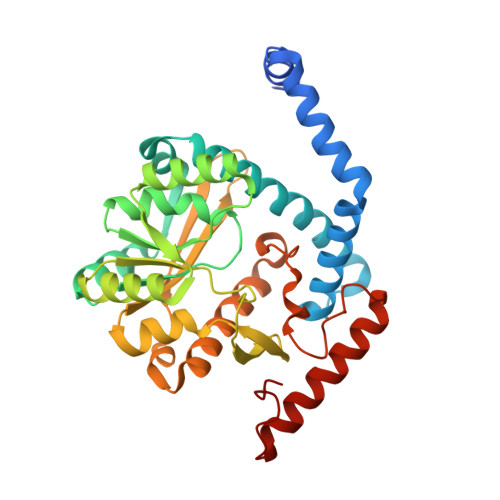
Structure-guided enzymology of the lipid A acyltransferase LpxM reveals a dual activity mechanism.

Dovala, D.Rath, C.M.Hu, Q.Sawyer, W.S.Shia, S.Elling, R.A.Knapp, M.S.Metzger, L.E.

(2016) Proc Natl Acad Sci U S A 113: E6064-E6071

  • DOI: https://doi.org/10.1073/pnas.1610746113
  • Primary Citation of Related Structures:  
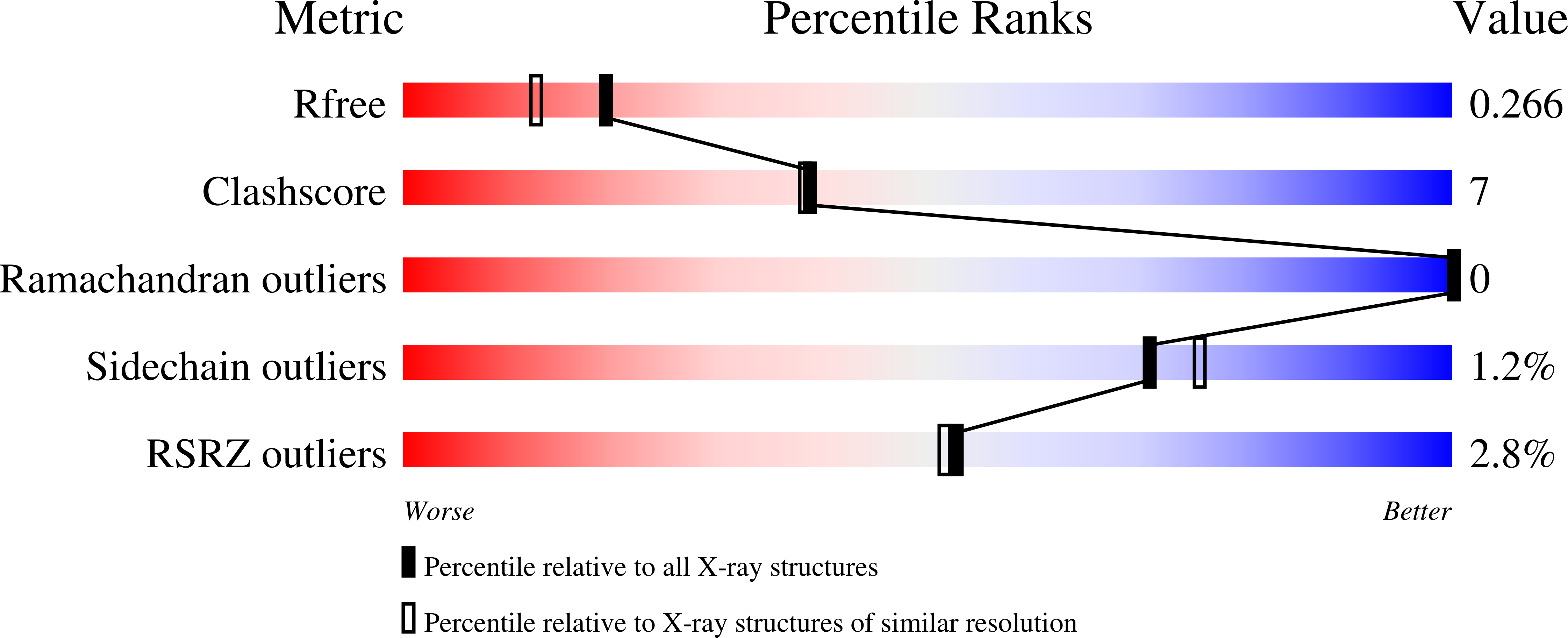
    5KN7, 5KNK

  • PubMed Abstract: 

    Gram-negative bacteria possess a characteristic outer membrane, of which the lipid A constituent elicits a strong host immune response through the Toll-like receptor 4 complex, and acts as a component of the permeability barrier to prevent uptake of bactericidal compounds. Lipid A species comprise the bulk of the outer leaflet of the outer membrane and are produced through a multistep biosynthetic pathway conserved in most Gram-negative bacteria. The final steps in this pathway involve the secondary acylation of lipid A precursors. These are catalyzed by members of a superfamily of enzymes known as lysophospholipid acyltransferases (LPLATs), which are present in all domains of life and play important roles in diverse biological processes. To date, characterization of this clinically important class of enzymes has been limited by a lack of structural information and the availability of only low-throughput biochemical assays. In this work, we present the structure of the bacterial LPLAT protein LpxM, and we describe a high-throughput, label-free mass spectrometric assay to characterize acyltransferase enzymatic activity. Using our structure and assay, we identify an LPLAT thioesterase activity, and we provide experimental evidence to support an ordered-binding and "reset" mechanistic model for LpxM function. This work enables the interrogation of other bacterial acyltransferases' structure-mechanism relationships, and the assay described herein provides a foundation for quantitatively characterizing the enzymology of any number of clinically relevant LPLAT proteins.


  • Organizational Affiliation

    Infectious Diseases, Novartis Institutes for BioMedical Research, Emeryville, CA 94608.


Macromolecules
Find similar proteins by:  (by identity cutoff)  |  3D Structure
Entity ID: 1
MoleculeChains Sequence LengthOrganismDetailsImage
Lipid A biosynthesis lauroyl acyltransferaseA [auth B]333Acinetobacter baumannii NIPH 410Mutation(s): 0 
Gene Names: F910_00940
Membrane Entity: Yes 
UniProt
Find proteins for S3TFW2 (Acinetobacter baumannii NIPH 410)
Explore S3TFW2 
Go to UniProtKB:  S3TFW2
Entity Groups  
Sequence Clusters30% Identity50% Identity70% Identity90% Identity95% Identity100% Identity
UniProt GroupS3TFW2
Sequence Annotations
Expand
  • Reference Sequence
Small Molecules
Ligands 5 Unique
IDChains Name / Formula / InChI Key2D Diagram3D Interactions
LMT
Query on LMT

Download Ideal Coordinates CCD File 
P [auth B]DODECYL-BETA-D-MALTOSIDE
C24 H46 O11
NLEBIOOXCVAHBD-QKMCSOCLSA-N
SO4
Query on SO4

Download Ideal Coordinates CCD File 
B,
C [auth B],
D [auth B]
SULFATE ION
O4 S
QAOWNCQODCNURD-UHFFFAOYSA-L
PO4
Query on PO4

Download Ideal Coordinates CCD File 
Q [auth B]PHOSPHATE ION
O4 P
NBIIXXVUZAFLBC-UHFFFAOYSA-K
GOL
Query on GOL

Download Ideal Coordinates CCD File 
H [auth B]
I [auth B]
J [auth B]
K [auth B]
L [auth B]
H [auth B],
I [auth B],
J [auth B],
K [auth B],
L [auth B],
M [auth B],
N [auth B],
O [auth B]
GLYCEROL
C3 H8 O3
PEDCQBHIVMGVHV-UHFFFAOYSA-N
NA
Query on NA

Download Ideal Coordinates CCD File 
E [auth B],
F [auth B],
G [auth B]
SODIUM ION
Na
FKNQFGJONOIPTF-UHFFFAOYSA-N
Experimental Data & Validation

Experimental Data

  • Method: X-RAY DIFFRACTION
  • Resolution: 1.99 Å
  • R-Value Free: 0.267 
  • R-Value Work: 0.213 
  • R-Value Observed: 0.215 
  • Space Group: C 2 2 21
Unit Cell:
Length ( Å )Angle ( ˚ )
a = 58.77α = 90
b = 145.33β = 90
c = 87.21γ = 90
Software Package:
Software NamePurpose
Aimlessdata scaling
PHENIXrefinement
PDB_EXTRACTdata extraction
iMOSFLMdata reduction
PHASERphasing

Structure Validation

View Full Validation Report


Ligand Structure Quality Assessment 


Entry History 

Deposition Data

Revision History  (Full details and data files)

  • Version 1.0: 2016-09-28
    Type: Initial release
  • Version 1.1: 2016-10-05
    Changes: Database references
  • Version 1.2: 2016-10-19
    Changes: Database references
  • Version 1.3: 2016-10-26
    Changes: Database references
  • Version 1.4: 2024-03-06
    Changes: Data collection, Database references, Derived calculations