


Crystal structure of enoyl-CoA hydratase from Mycobacterium tuberculosis H37Rv
Nocek, B., Hatzos-Skintges, C., Anderson, W.F., Joachimiak, A., Center for Structural Genomics of Infectious Diseases (CSGID)To be published.
Experimental Data Snapshot
wwPDB Validation 3D Report Full Report

Entity ID: 1 | |||||
|---|---|---|---|---|---|
| Molecule | Chains | Sequence Length | Organism | Details | Image |
| Enoyl-CoA hydratase | 265 | Mycobacterium tuberculosis H37Rv | Mutation(s): 0 Gene Names: echA1, Rv0222, LH57_01220 EC: 4.2.1.17 | 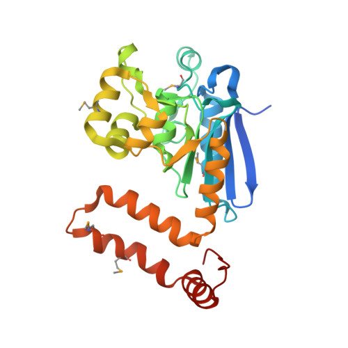 | |
UniProt | |||||
Find proteins for P96404 (Mycobacterium tuberculosis (strain ATCC 25618 / H37Rv)) Explore P96404 Go to UniProtKB: P96404 | |||||
Entity Groups | |||||
| Sequence Clusters | 30% Identity50% Identity70% Identity90% Identity95% Identity100% Identity | ||||
| UniProt Group | P96404 | ||||
Sequence AnnotationsExpand | |||||
| |||||
| Modified Residues 1 Unique | |||||
|---|---|---|---|---|---|
| ID | Chains | Type | Formula | 2D Diagram | Parent |
| MSE Query on MSE | A | L-PEPTIDE LINKING | C5 H11 N O2 Se |  | MET |
| Length ( Å ) | Angle ( ˚ ) |
|---|---|
| a = 121.476 | α = 90 |
| b = 121.476 | β = 90 |
| c = 121.476 | γ = 90 |
| Software Name | Purpose |
|---|---|
| PHENIX | refinement |
| HKL-3000 | data reduction |
| HKL-3000 | data scaling |
| SHELXDE | phasing |
RCSB PDB (citation) is hosted by
RCSB PDB is a member of the

