5KHE

Fitted structure of rubella virus capsid protein


Experimental Data Snapshot

  • Method: ELECTRON MICROSCOPY
  • Resolution: 35.0 Å
  • Aggregation State: PARTICLE 
  • Reconstruction Method: SUBTOMOGRAM AVERAGING 

wwPDB Validation   3D Report Full Report


This is version 1.3 of the entry. See complete history


Literature

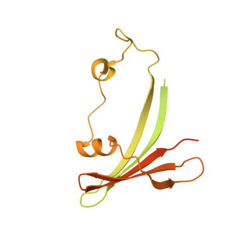
Assembly, maturation and three-dimensional helical structure of the teratogenic rubella virus.

Mangala Prasad, V.Klose, T.Rossmann, M.G.

(2017) PLoS Pathog 13: e1006377-e1006377

  • DOI: https://doi.org/10.1371/journal.ppat.1006377
  • Primary Citation of Related Structures:  
    5KHC, 5KHE, 5KHF

  • PubMed Abstract: 

    Viral infections during pregnancy are a significant cause of infant morbidity and mortality. Of these, rubella virus infection is a well-substantiated example that leads to miscarriages or severe fetal defects. However, structural information about the rubella virus has been lacking due to the pleomorphic nature of the virions. Here we report a helical structure of rubella virions using cryo-electron tomography. Sub-tomogram averaging of the surface spikes established the relative positions of the viral glycoproteins, which differed from the earlier icosahedral models of the virus. Tomographic analyses of in vitro assembled nucleocapsids and virions provide a template for viral assembly. Comparisons of immature and mature virions show large rearrangements in the glycoproteins that may be essential for forming the infectious virions. These results present the first known example of a helical membrane-enveloped virus, while also providing a structural basis for its assembly and maturation pathway.


  • Organizational Affiliation

    Department of Biological Sciences, 240 S. Martin Jischke Drive, Purdue University, West Lafayette, IN, United States of America.


Macromolecules
Find similar proteins by:  (by identity cutoff)  |  3D Structure
Entity ID: 1
MoleculeChains Sequence LengthOrganismDetailsImage
capsid protein
A, B
269Rubella virusMutation(s): 0 
UniProt
Find proteins for P07566 (Rubella virus (strain Therien))
Explore P07566 
Go to UniProtKB:  P07566
Entity Groups  
Sequence Clusters30% Identity50% Identity70% Identity90% Identity95% Identity100% Identity
UniProt GroupP07566
Sequence Annotations
Expand
  • Reference Sequence
Experimental Data & Validation

Experimental Data

  • Method: ELECTRON MICROSCOPY
  • Resolution: 35.0 Å
  • Aggregation State: PARTICLE 
  • Reconstruction Method: SUBTOMOGRAM AVERAGING 
EM Software:
TaskSoftware PackageVersion
RECONSTRUCTIONPEET10.1

Structure Validation

View Full Validation Report


Entry History & Funding Information

Deposition Data


Funding OrganizationLocationGrant Number
National Institutes of Health/National Institute Of Allergy and Infectious Diseases (NIH/NIAID)United StatesAI095366

Revision History  (Full details and data files)

  • Version 1.0: 2017-05-10
    Type: Initial release
  • Version 1.1: 2017-06-14
    Changes: Database references
  • Version 1.2: 2017-09-13
    Changes: Author supporting evidence, Data collection
  • Version 1.3: 2019-12-11
    Changes: Author supporting evidence