5K21

Pyocyanin demethylase


Experimental Data Snapshot

  • Method: X-RAY DIFFRACTION
  • Resolution: 1.80 Å
  • R-Value Free: 0.195 
  • R-Value Work: 0.168 

wwPDB Validation   3D Report Full Report


Ligand Structure Quality Assessment 


This is version 1.4 of the entry. See complete history


Literature

Pyocyanin degradation by a tautomerizing demethylase inhibits Pseudomonas aeruginosa biofilms.

Costa, K.C.Glasser, N.R.Conway, S.J.Newman, D.K.

(2017) Science 355: 170-173

  • DOI: https://doi.org/10.1126/science.aag3180
  • Primary Citation of Related Structures:  
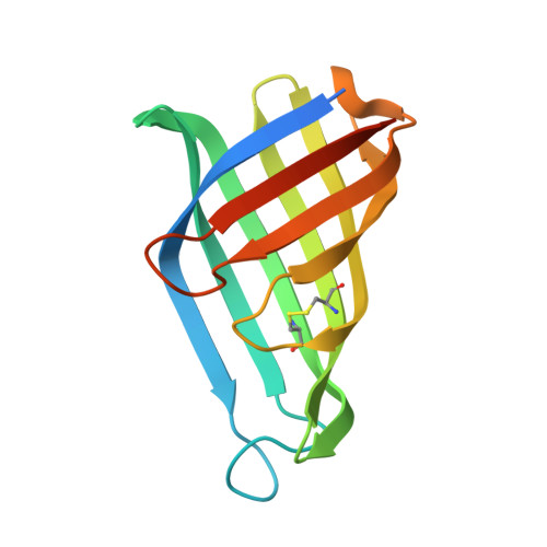
    5K21

  • PubMed Abstract: 

    The opportunistic pathogen Pseudomonas aeruginosa produces colorful redox-active metabolites called phenazines, which underpin biofilm development, virulence, and clinical outcomes. Although phenazines exist in many forms, the best studied is pyocyanin. Here, we describe pyocyanin demethylase (PodA), a hitherto uncharacterized protein that oxidizes the pyocyanin methyl group to formaldehyde and reduces the pyrazine ring via an unusual tautomerizing demethylation reaction. Treatment with PodA disrupts P. aeruginosa biofilm formation similarly to DNase, suggesting interference with the pyocyanin-dependent release of extracellular DNA into the matrix. PodA-dependent pyocyanin demethylation also restricts established biofilm aggregate populations experiencing anoxic conditions. Together, these results show that modulating extracellular redox-active metabolites can influence the fitness of a biofilm-forming microorganism.


  • Organizational Affiliation

    Division of Biology and Biological Engineering, California Institute of Technology, Pasadena, CA 91125, USA.


Macromolecules
Find similar proteins by:  (by identity cutoff)  |  3D Structure
Entity ID: 1
MoleculeChains Sequence LengthOrganismDetailsImage
Pyocyanin demethylase
A, B, C
141Mycolicibacterium fortuitum subsp. fortuitum DSM 46621 = ATCC 6841 = JCM 6387Mutation(s): 0 
Gene Names: MFORT_14352
UniProt
Find proteins for K0V2D8 (Mycolicibacterium fortuitum subsp. fortuitum DSM 46621 = ATCC 6841 = JCM 6387)
Explore K0V2D8 
Go to UniProtKB:  K0V2D8
Entity Groups  
Sequence Clusters30% Identity50% Identity70% Identity90% Identity95% Identity100% Identity
UniProt GroupK0V2D8
Sequence Annotations
Expand
  • Reference Sequence
Experimental Data & Validation

Experimental Data

  • Method: X-RAY DIFFRACTION
  • Resolution: 1.80 Å
  • R-Value Free: 0.195 
  • R-Value Work: 0.168 
  • Space Group: P 21 21 21
Unit Cell:
Length ( Å )Angle ( ˚ )
a = 65.33α = 90
b = 72.98β = 90
c = 79.7γ = 90
Software Package:
Software NamePurpose
PHENIXrefinement
XDSdata scaling
XSCALEdata scaling
PHENIXmodel building
PHASERphasing

Structure Validation

View Full Validation Report


Ligand Structure Quality Assessment 


Entry History & Funding Information

Deposition Data


Funding OrganizationLocationGrant Number
Howard Hughes Medical Institute (HHMI)United States--
National Institutes of Health/National Institute Of Allergy and Infectious Diseases (NIH/NIAID)United StatesF32AI112248
National Institutes of Health/National Heart, Lung, and Blood Institute (NIH/NHLBI)United States5R01HL117328-03

Revision History  (Full details and data files)

  • Version 1.0: 2016-12-07
    Type: Initial release
  • Version 1.1: 2017-01-11
    Changes: Database references
  • Version 1.2: 2017-01-25
    Changes: Database references
  • Version 1.3: 2017-09-20
    Changes: Author supporting evidence, Refinement description
  • Version 1.4: 2019-11-20
    Changes: Author supporting evidence