5JJC

Crystal Structure of double mutant (Q96A-Y125A) O-Acetyl Serine Sulfhydralase from Brucella abortus


Experimental Data Snapshot

  • Method: X-RAY DIFFRACTION
  • Resolution: 2.01 Å
  • R-Value Free: 0.208 
  • R-Value Work: 0.168 
  • R-Value Observed: 0.170 

wwPDB Validation   3D Report Full Report


This is version 1.2 of the entry. See complete history


Literature

Structure-based mutational studies of O-acetylserine sulfhydrylase reveal the reason for the loss of cysteine synthase complex formation in Brucella abortus

Dharavath, S.Raj, I.Gourinath, S.

(2017) Biochem J 474: 1221-1239

  • DOI: https://doi.org/10.1042/BCJ20161062
  • Primary Citation of Related Structures:  
    5JIS, 5JJC

  • PubMed Abstract: 

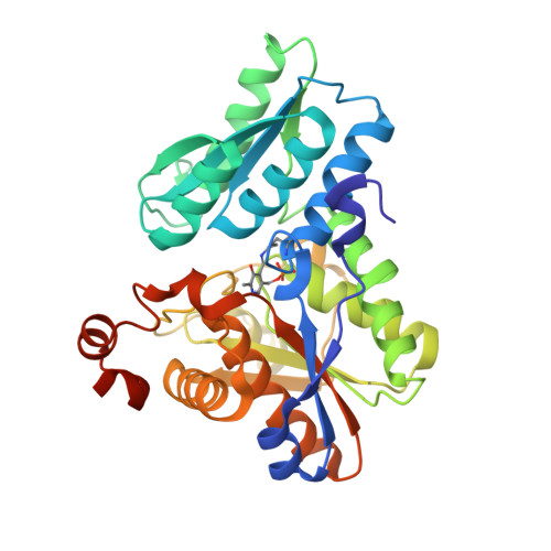
    Cysteine biosynthesis takes place via a two-step pathway in bacteria, fungi, plants and protozoan parasites, but not in humans, and hence, the machinery of cysteine biosynthesis is an opportune target for therapeutics. The decameric cysteine synthase complex (CSC) is formed when the C-terminal tail of serine acetyltransferase (SAT) binds in the active site of O -acetylserine sulfydrylase (OASS), playing a role in the regulation of this pathway. Here, we show that OASS from Brucella abortus (BaOASS) does not interact with its cognate SAT C-terminal tail. Crystal structures of native BaOASS showed that residues Gln96 and Tyr125 occupy the active-site pocket and interfere with the entry of the SAT C-terminal tail. The BaOASS (Q96A-Y125A) mutant showed relatively strong binding ( K d  = 32.4 μM) to BaSAT C-terminal peptides in comparison with native BaOASS. The mutant structure looks similar except that the active-site pocket has enough space to bind the SAT C-terminal end. Surface plasmon resonance results showed a relatively strong (7.3 μM K d ) interaction between BaSAT and the BaOASS (Q96A-Y125A), but no interaction with native BaOASS. Taken together, our observations suggest that the CSC does not form in B. abortus .


  • Organizational Affiliation

    School of Life Sciences, Jawaharlal Nehru University, New Delhi - 110067, India.


Macromolecules
Find similar proteins by:  (by identity cutoff)  |  3D Structure
Entity ID: 1
MoleculeChains Sequence LengthOrganismDetailsImage
Cysteine synthase
A, B, C, D
342Brucella abortus S19Mutation(s): 2 
Gene Names: BAbS19_I09950
EC: 2.5.1.47
UniProt
Find proteins for A0A0F6AQU1 (Brucella abortus (strain S19))
Explore A0A0F6AQU1 
Go to UniProtKB:  A0A0F6AQU1
Entity Groups  
Sequence Clusters30% Identity50% Identity70% Identity90% Identity95% Identity100% Identity
UniProt GroupA0A0F6AQU1
Sequence Annotations
Expand
  • Reference Sequence
Small Molecules
Modified Residues  1 Unique
IDChains TypeFormula2D DiagramParent
LLP
Query on LLP
A, B, C, D
L-PEPTIDE LINKINGC14 H22 N3 O7 PLYS
Experimental Data & Validation

Experimental Data

  • Method: X-RAY DIFFRACTION
  • Resolution: 2.01 Å
  • R-Value Free: 0.208 
  • R-Value Work: 0.168 
  • R-Value Observed: 0.170 
  • Space Group: P 61
Unit Cell:
Length ( Å )Angle ( ˚ )
a = 104.82α = 90
b = 104.82β = 90
c = 242.041γ = 120
Software Package:
Software NamePurpose
PHENIXrefinement
HKL-2000data reduction
HKL-2000data scaling
PHASERphasing

Structure Validation

View Full Validation Report


Entry History 

Deposition Data

Revision History  (Full details and data files)

  • Version 1.0: 2017-04-05
    Type: Initial release
  • Version 1.1: 2023-11-08
    Changes: Data collection, Database references, Refinement description
  • Version 1.2: 2023-11-15
    Changes: Data collection