5J2M

HIV-1 reverse transcriptase in complex with DNA and EFdA-triphosphate, a translocation-defective RT inhibitor


Experimental Data Snapshot

  • Method: X-RAY DIFFRACTION
  • Resolution: 2.43 Å
  • R-Value Free: 0.233 
  • R-Value Work: 0.199 
  • R-Value Observed: 0.201 

wwPDB Validation   3D Report Full Report


This is version 1.5 of the entry. See complete history


Literature

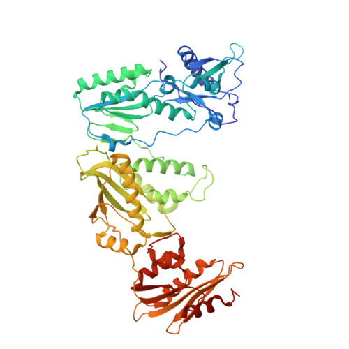
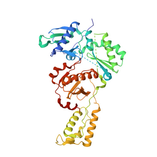
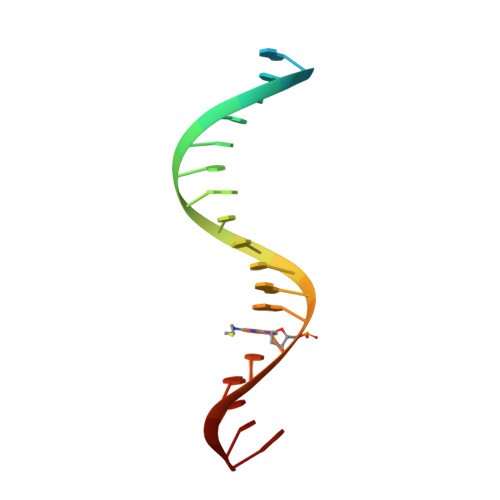
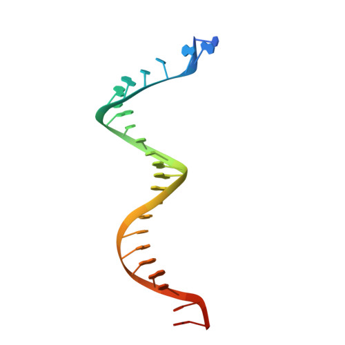
Structural basis of HIV inhibition by translocation-defective RT inhibitor 4'-ethynyl-2-fluoro-2'-deoxyadenosine (EFdA).

Salie, Z.L.Kirby, K.A.Michailidis, E.Marchand, B.Singh, K.Rohan, L.C.Kodama, E.N.Mitsuya, H.Parniak, M.A.Sarafianos, S.G.

(2016) Proc Natl Acad Sci U S A 113: 9274-9279

  • DOI: https://doi.org/10.1073/pnas.1605223113
  • Primary Citation of Related Structures:  
    5J2M, 5J2N, 5J2P, 5J2Q

  • PubMed Abstract: 

    4'-Ethynyl-2-fluoro-2'-deoxyadenosine (EFdA) is the most potent nucleoside analog inhibitor of HIV reverse transcriptase (RT). It retains a 3'-OH yet acts as a chain-terminating agent by diminishing translocation from the pretranslocation nucleotide-binding site (N site) to the posttranslocation primer-binding site (P site). Also, facile misincorporation of EFdA-monophosphate (MP) results in difficult-to-extend mismatched primers. To understand the high potency and unusual inhibition mechanism of EFdA, we solved RT crystal structures (resolutions from 2.4 to 2.9 Å) that include inhibition intermediates (i) before inhibitor incorporation (catalytic complex, RT/DNA/EFdA-triphosphate), (ii) after incorporation of EFdA-MP followed by dT-MP (RT/DNAEFdA-MP(P)• dT-MP(N) ), or (iii) after incorporation of two EFdA-MPs (RT/DNAEFdA-MP(P)• EFdA-MP(N) ); (iv) the latter was also solved with EFdA-MP mismatched at the N site (RT/DNAEFdA-MP(P)• EFdA-MP(*N) ). We report that the inhibition mechanism and potency of EFdA stem from interactions of its 4'-ethynyl at a previously unexploited conserved hydrophobic pocket in the polymerase active site. The high resolution of the catalytic complex structure revealed a network of ordered water molecules at the polymerase active site that stabilize enzyme interactions with nucleotide and DNA substrates. Finally, decreased translocation results from favorable interactions of primer-terminating EFdA-MP at the pretranslocation site and unfavorable posttranslocation interactions that lead to observed localized primer distortions.


  • Organizational Affiliation

    C. S. Bond Life Sciences Center, Department of Biochemistry, University of Missouri, Columbia, MO 65211;


Macromolecules

Find similar proteins by:  (by identity cutoff)  |  3D Structure
Entity ID: 1
MoleculeChains Sequence LengthOrganismDetailsImage
HIV-1 reverse transcriptase p51 subunit560HIV-1 M:B_HXB2RMutation(s): 2 
Gene Names: gag-pol
EC: 2.7.7
UniProt
Find proteins for P04585 (Human immunodeficiency virus type 1 group M subtype B (isolate HXB2))
Explore P04585 
Go to UniProtKB:  P04585
Entity Groups  
Sequence Clusters30% Identity50% Identity70% Identity90% Identity95% Identity100% Identity
UniProt GroupP04585
Sequence Annotations
Expand
  • Reference Sequence
Find similar proteins by:  (by identity cutoff)  |  3D Structure
Entity ID: 2
MoleculeChains Sequence LengthOrganismDetailsImage
HIV-1 reverse transcriptase p51 subunit440HIV-1 M:B_HXB2RMutation(s): 1 
Gene Names: gag-pol
EC: 2.7.7
UniProt
Find proteins for P04585 (Human immunodeficiency virus type 1 group M subtype B (isolate HXB2))
Explore P04585 
Go to UniProtKB:  P04585
Entity Groups  
Sequence Clusters30% Identity50% Identity70% Identity90% Identity95% Identity100% Identity
UniProt GroupP04585
Sequence Annotations
Expand
  • Reference Sequence

Find similar nucleic acids by:  Sequence   |   3D Structure  

Entity ID: 3
MoleculeChains LengthOrganismImage
DNA (5'-D(*AP*CP*AP*GP*TP*CP*CP*CP*TP*GP*TP*TP*CP*GP*GP*(MRG)P*CP*GP*CP*CP*G)-3')C [auth P]21Human immunodeficiency virus 1
Sequence Annotations
Expand
  • Reference Sequence
Find similar nucleic acids by:  (by identity cutoff)  |  3D Structure
Entity ID: 4
MoleculeChains LengthOrganismImage
DNA (27-MER)D [auth T]27Human immunodeficiency virus 1
Sequence Annotations
Expand
  • Reference Sequence
Experimental Data & Validation

Experimental Data

  • Method: X-RAY DIFFRACTION
  • Resolution: 2.43 Å
  • R-Value Free: 0.233 
  • R-Value Work: 0.199 
  • R-Value Observed: 0.201 
  • Space Group: C 2 2 21
Unit Cell:
Length ( Å )Angle ( ˚ )
a = 167.468α = 90
b = 169.758β = 90
c = 101.951γ = 90
Software Package:
Software NamePurpose
PHENIXrefinement
XDSdata reduction
Aimlessdata scaling
PHASERphasing
PDB_EXTRACTdata extraction

Structure Validation

View Full Validation Report


Entry History & Funding Information

Deposition Data


Funding OrganizationLocationGrant Number
National Institutes of Health/National Institute Of Allergy and Infectious Diseases (NIH/NIAID)United StatesAI076119
National Institutes of Health/National Institute Of Allergy and Infectious Diseases (NIH/NIAID)United StatesAI099284
National Institutes of Health/National Institute Of Allergy and Infectious Diseases (NIH/NIAID)United StatesAI100890
National Institutes of Health/National Institute Of Allergy and Infectious Diseases (NIH/NIAID)United StatesAI112417
National Institutes of Health/National Institute Of Allergy and Infectious Diseases (NIH/NIAID)United StatesAI120860
National Institutes of Health/National Institute of General Medical Sciences (NIH/NIGMS)United StatesGM103368
University of MissouriUnited StatesMizzou Advantage

Revision History  (Full details and data files)

  • Version 1.0: 2016-08-03
    Type: Initial release
  • Version 1.1: 2016-08-17
    Changes: Database references
  • Version 1.2: 2016-08-31
    Changes: Database references
  • Version 1.3: 2017-09-20
    Changes: Advisory, Author supporting evidence, Database references, Derived calculations
  • Version 1.4: 2019-12-11
    Changes: Author supporting evidence
  • Version 1.5: 2023-09-27
    Changes: Advisory, Data collection, Database references, Derived calculations, Refinement description