5I85

aSMase with zinc and phosphocholine


Experimental Data Snapshot

  • Method: X-RAY DIFFRACTION
  • Resolution: 2.50 Å
  • R-Value Free: 0.222 
  • R-Value Work: 0.190 
  • R-Value Observed: 0.192 

wwPDB Validation   3D Report Full Report


Ligand Structure Quality Assessment 


This is version 2.2 of the entry. See complete history


Literature

Human acid sphingomyelinase structures provide insight to molecular basis of Niemann-Pick disease.

Zhou, Y.F.Metcalf, M.C.Garman, S.C.Edmunds, T.Qiu, H.Wei, R.R.

(2016) Nat Commun 7: 13082-13082

  • DOI: https://doi.org/10.1038/ncomms13082
  • Primary Citation of Related Structures:  
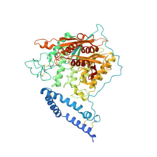
    5I81, 5I85, 5I8R

  • PubMed Abstract: 

    Acid sphingomyelinase (ASM) hydrolyzes sphingomyelin to ceramide and phosphocholine, essential components of myelin in neurons. Genetic alterations in ASM lead to ASM deficiency (ASMD) and have been linked to Niemann-Pick disease types A and B. Olipudase alfa, a recombinant form of human ASM, is being developed as enzyme replacement therapy to treat the non-neurological manifestations of ASMD. Here we present the human ASM holoenzyme and product bound structures encompassing all of the functional domains. The catalytic domain has a metallophosphatase fold, and two zinc ions and one reaction product phosphocholine are identified in a histidine-rich active site. The structures reveal the underlying catalytic mechanism, in which two zinc ions activate a water molecule for nucleophilic attack of the phosphodiester bond. Docking of sphingomyelin provides a model that allows insight into the selectivity of the enzyme and how the ASM domains collaborate to complete hydrolysis. Mapping of known mutations provides a basic understanding on correlations between enzyme dysfunction and phenotypes observed in ASMD patients.


  • Organizational Affiliation

    Protein Engineering Department, Biologics Research, Sanofi, Framingham, Massachusetts 01701, USA.


Macromolecules
Find similar proteins by:  (by identity cutoff)  |  3D Structure
Entity ID: 1
MoleculeChains Sequence LengthOrganismDetailsImage
Sphingomyelin phosphodiesterase583Homo sapiensMutation(s): 0 
Gene Names: SMPD1ASM
EC: 3.1.4.12
Membrane Entity: Yes 
UniProt & NIH Common Fund Data Resources
Find proteins for P17405 (Homo sapiens)
Explore P17405 
Go to UniProtKB:  P17405
PHAROS:  P17405
GTEx:  ENSG00000166311 
Entity Groups  
Sequence Clusters30% Identity50% Identity70% Identity90% Identity95% Identity100% Identity
UniProt GroupP17405
Sequence Annotations
Expand
  • Reference Sequence
Oligosaccharides

Help

Entity ID: 2
MoleculeChains Length2D Diagram Glycosylation3D Interactions
beta-D-mannopyranose-(1-4)-2-acetamido-2-deoxy-beta-D-glucopyranose-(1-4)-2-acetamido-2-deoxy-beta-D-glucopyranose
B
3N/A
Glycosylation Resources
GlyTouCan:  G15407YE
GlyCosmos:  G15407YE
GlyGen:  G15407YE
Entity ID: 3
MoleculeChains Length2D Diagram Glycosylation3D Interactions
alpha-D-mannopyranose-(1-3)-alpha-D-mannopyranose-(1-6)-beta-D-mannopyranose-(1-4)-2-acetamido-2-deoxy-beta-D-glucopyranose-(1-4)-2-acetamido-2-deoxy-beta-D-glucopyranose
C
5N-Glycosylation
Glycosylation Resources
GlyTouCan:  G10756ZZ
GlyCosmos:  G10756ZZ
GlyGen:  G10756ZZ
Entity ID: 4
MoleculeChains Length2D Diagram Glycosylation3D Interactions
2-acetamido-2-deoxy-beta-D-glucopyranose-(1-4)-2-acetamido-2-deoxy-beta-D-glucopyranose
D
2N-Glycosylation
Glycosylation Resources
GlyTouCan:  G42666HT
GlyCosmos:  G42666HT
GlyGen:  G42666HT
Small Molecules
Ligands 4 Unique
IDChains Name / Formula / InChI Key2D Diagram3D Interactions
NAG
Query on NAG

Download Ideal Coordinates CCD File 
E [auth A],
F [auth A],
G [auth A]
2-acetamido-2-deoxy-beta-D-glucopyranose
C8 H15 N O6
OVRNDRQMDRJTHS-FMDGEEDCSA-N
PC
Query on PC

Download Ideal Coordinates CCD File 
U [auth A]PHOSPHOCHOLINE
C5 H15 N O4 P
YHHSONZFOIEMCP-UHFFFAOYSA-O
SO4
Query on SO4

Download Ideal Coordinates CCD File 
J [auth A]
K [auth A]
L [auth A]
M [auth A]
N [auth A]
J [auth A],
K [auth A],
L [auth A],
M [auth A],
N [auth A],
O [auth A],
P [auth A],
Q [auth A],
R [auth A],
S [auth A],
T [auth A]
SULFATE ION
O4 S
QAOWNCQODCNURD-UHFFFAOYSA-L
ZN
Query on ZN

Download Ideal Coordinates CCD File 
H [auth A],
I [auth A]
ZINC ION
Zn
PTFCDOFLOPIGGS-UHFFFAOYSA-N
Experimental Data & Validation

Experimental Data

  • Method: X-RAY DIFFRACTION
  • Resolution: 2.50 Å
  • R-Value Free: 0.222 
  • R-Value Work: 0.190 
  • R-Value Observed: 0.192 
  • Space Group: P 64 2 2
Unit Cell:
Length ( Å )Angle ( ˚ )
a = 131.601α = 90
b = 131.601β = 90
c = 188.573γ = 120
Software Package:
Software NamePurpose
PHENIXrefinement
CrystalCleardata collection
SCALAdata scaling
MOLREPphasing
PDB_EXTRACTdata extraction
iMOSFLMdata reduction

Structure Validation

View Full Validation Report


Ligand Structure Quality Assessment 


Entry History 

Deposition Data

Revision History  (Full details and data files)

  • Version 1.0: 2016-09-07
    Type: Initial release
  • Version 1.1: 2016-11-16
    Changes: Database references
  • Version 2.0: 2020-07-29
    Type: Remediation
    Reason: Carbohydrate remediation
    Changes: Advisory, Atomic model, Data collection, Derived calculations, Structure summary
  • Version 2.1: 2021-03-24
    Changes: Derived calculations, Source and taxonomy, Structure summary
  • Version 2.2: 2023-09-27
    Changes: Data collection, Database references, Refinement description