5HKK

Caldalaklibacillus thermarum F1-ATPase (wild type)


Experimental Data Snapshot

  • Method: X-RAY DIFFRACTION
  • Resolution: 3.00 Å
  • R-Value Free: 0.246 
  • R-Value Work: 0.203 
  • R-Value Observed: 0.205 

wwPDB Validation   3D Report Full Report


Ligand Structure Quality Assessment 


This is version 1.4 of the entry. See complete history


Literature

Regulation of the thermoalkaliphilic F1-ATPase from Caldalkalibacillus thermarum.

Ferguson, S.A.Cook, G.M.Montgomery, M.G.Leslie, A.G.Walker, J.E.

(2016) Proc Natl Acad Sci U S A 113: 10860-10865

  • DOI: https://doi.org/10.1073/pnas.1612035113
  • Primary Citation of Related Structures:  
    5HKK, 5IK2

  • PubMed Abstract: 

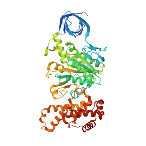
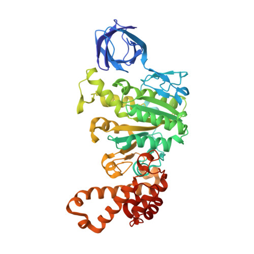
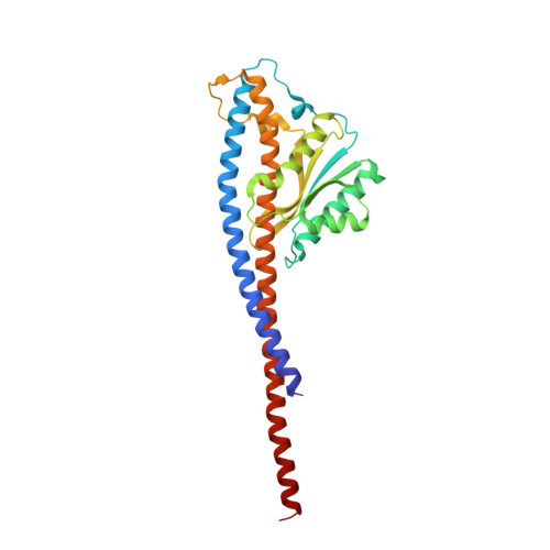
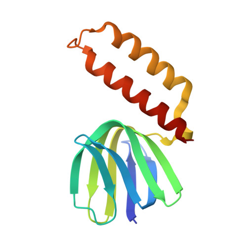
    The crystal structure has been determined of the F1-catalytic domain of the F-ATPase from Caldalkalibacillus thermarum, which hydrolyzes adenosine triphosphate (ATP) poorly. It is very similar to those of active mitochondrial and bacterial F1-ATPases. In the F-ATPase from Geobacillus stearothermophilus, conformational changes in the ε-subunit are influenced by intracellular ATP concentration and membrane potential. When ATP is plentiful, the ε-subunit assumes a "down" state, with an ATP molecule bound to its two C-terminal α-helices; when ATP is scarce, the α-helices are proposed to inhibit ATP hydrolysis by assuming an "up" state, where the α-helices, devoid of ATP, enter the α3β3-catalytic region. However, in the Escherichia coli enzyme, there is no evidence that such ATP binding to the ε-subunit is mechanistically important for modulating the enzyme's hydrolytic activity. In the structure of the F1-ATPase from C. thermarum, ATP and a magnesium ion are bound to the α-helices in the down state. In a form with a mutated ε-subunit unable to bind ATP, the enzyme remains inactive and the ε-subunit is down. Therefore, neither the γ-subunit nor the regulatory ATP bound to the ε-subunit is involved in the inhibitory mechanism of this particular enzyme. The structure of the α3β3-catalytic domain is likewise closely similar to those of active F1-ATPases. However, although the βE-catalytic site is in the usual "open" conformation, it is occupied by the unique combination of an ADP molecule with no magnesium ion and a phosphate ion. These bound hydrolytic products are likely to be the basis of inhibition of ATP hydrolysis.


  • Organizational Affiliation

    Medical Research Council Mitochondrial Biology Unit, Cambridge Biomedical Campus, Cambridge CB2 0XY, United Kingdom;


Macromolecules
Find similar proteins by:  (by identity cutoff)  |  3D Structure
Entity ID: 1
MoleculeChains Sequence LengthOrganismDetailsImage
ATP synthase subunit alpha
A, B, C, I, J
A, B, C, I, J, K
502Caldalkalibacillus thermarum TA2.A1Mutation(s): 0 
Gene Names: atpACathTA2_2809
EC: 3.6.3.14
Membrane Entity: Yes 
UniProt
Find proteins for F5LA74 (Caldalkalibacillus thermarum (strain TA2.A1))
Explore F5LA74 
Go to UniProtKB:  F5LA74
Entity Groups  
Sequence Clusters30% Identity50% Identity70% Identity90% Identity95% Identity100% Identity
UniProt GroupF5LA74
Sequence Annotations
Expand
  • Reference Sequence
Find similar proteins by:  (by identity cutoff)  |  3D Structure
Entity ID: 2
MoleculeChains Sequence LengthOrganismDetailsImage
ATP synthase subunit beta
D, E, F, L, M
D, E, F, L, M, N
462Caldalkalibacillus thermarum TA2.A1Mutation(s): 0 
Gene Names: atpDCathTA2_2807
EC: 3.6.3.14
Membrane Entity: Yes 
UniProt
Find proteins for F5LA72 (Caldalkalibacillus thermarum (strain TA2.A1))
Explore F5LA72 
Go to UniProtKB:  F5LA72
Entity Groups  
Sequence Clusters30% Identity50% Identity70% Identity90% Identity95% Identity100% Identity
UniProt GroupF5LA72
Sequence Annotations
Expand
  • Reference Sequence
Find similar proteins by:  (by identity cutoff)  |  3D Structure
Entity ID: 3
MoleculeChains Sequence LengthOrganismDetailsImage
ATP synthase gamma chain
G, O
286Caldalkalibacillus thermarum TA2.A1Mutation(s): 0 
Gene Names: atpGCathTA2_2808
Membrane Entity: Yes 
UniProt
Find proteins for F5LA73 (Caldalkalibacillus thermarum (strain TA2.A1))
Explore F5LA73 
Go to UniProtKB:  F5LA73
Entity Groups  
Sequence Clusters30% Identity50% Identity70% Identity90% Identity95% Identity100% Identity
UniProt GroupF5LA73
Sequence Annotations
Expand
  • Reference Sequence
Find similar proteins by:  (by identity cutoff)  |  3D Structure
Entity ID: 4
MoleculeChains Sequence LengthOrganismDetailsImage
ATP synthase epsilon chain
H, P
135Caldalkalibacillus thermarum TA2.A1Mutation(s): 0 
Gene Names: atpCCathTA2_2806
Membrane Entity: Yes 
UniProt
Find proteins for F5LA71 (Caldalkalibacillus thermarum (strain TA2.A1))
Explore F5LA71 
Go to UniProtKB:  F5LA71
Entity Groups  
Sequence Clusters30% Identity50% Identity70% Identity90% Identity95% Identity100% Identity
UniProt GroupF5LA71
Sequence Annotations
Expand
  • Reference Sequence
Small Molecules
Ligands 5 Unique
IDChains Name / Formula / InChI Key2D Diagram3D Interactions
ATP
Query on ATP

Download Ideal Coordinates CCD File 
EA [auth H],
SA [auth P]
ADENOSINE-5'-TRIPHOSPHATE
C10 H16 N5 O13 P3
ZKHQWZAMYRWXGA-KQYNXXCUSA-N
ADP
Query on ADP

Download Ideal Coordinates CCD File 
BA [auth F]
FA [auth I]
HA [auth J]
JA [auth K]
LA [auth L]
BA [auth F],
FA [auth I],
HA [auth J],
JA [auth K],
LA [auth L],
NA [auth M],
PA [auth N],
Q [auth A],
T [auth B],
V [auth C],
X [auth D],
Z [auth E]
ADENOSINE-5'-DIPHOSPHATE
C10 H15 N5 O10 P2
XTWYTFMLZFPYCI-KQYNXXCUSA-N
PO4
Query on PO4

Download Ideal Coordinates CCD File 
AA [auth E],
OA [auth M]
PHOSPHATE ION
O4 P
NBIIXXVUZAFLBC-UHFFFAOYSA-K
GOL
Query on GOL

Download Ideal Coordinates CCD File 
S [auth A]GLYCEROL
C3 H8 O3
PEDCQBHIVMGVHV-UHFFFAOYSA-N
MG
Query on MG

Download Ideal Coordinates CCD File 
CA [auth F]
DA [auth G]
GA [auth I]
IA [auth J]
KA [auth K]
CA [auth F],
DA [auth G],
GA [auth I],
IA [auth J],
KA [auth K],
MA [auth L],
QA [auth N],
R [auth A],
RA [auth O],
U [auth B],
W [auth C],
Y [auth D]
MAGNESIUM ION
Mg
JLVVSXFLKOJNIY-UHFFFAOYSA-N
Experimental Data & Validation

Experimental Data

  • Method: X-RAY DIFFRACTION
  • Resolution: 3.00 Å
  • R-Value Free: 0.246 
  • R-Value Work: 0.203 
  • R-Value Observed: 0.205 
  • Space Group: P 1 21 1
Unit Cell:
Length ( Å )Angle ( ˚ )
a = 147.82α = 90
b = 130.79β = 107.97
c = 210.39γ = 90
Software Package:
Software NamePurpose
MOSFLMdata reduction
Aimlessdata scaling
PHASERphasing
Cootmodel building
REFMACrefinement
PDB_EXTRACTdata extraction

Structure Validation

View Full Validation Report


Ligand Structure Quality Assessment 


Entry History & Funding Information

Deposition Data


Funding OrganizationLocationGrant Number
Medical Research Council (United Kingdom)United KingdomU1065663150
Medical Research Council (United Kingdom)United KingdomU105184325
Medical Research Council (United Kingdom)United KingdomMR/M009858/1
James Cook FellowshipNew Zealand--
Health Research CouncilNew Zealand--

Revision History  (Full details and data files)

  • Version 1.0: 2016-09-21
    Type: Initial release
  • Version 1.1: 2016-09-28
    Changes: Database references
  • Version 1.2: 2016-10-12
    Changes: Database references
  • Version 1.3: 2017-09-13
    Changes: Author supporting evidence
  • Version 1.4: 2024-01-10
    Changes: Data collection, Database references, Derived calculations, Refinement description