5HI9

Structure of the full-length TRPV2 channel by cryo-electron microscopy


Experimental Data Snapshot

  • Method: ELECTRON MICROSCOPY
  • Resolution: 4.40 Å
  • Aggregation State: PARTICLE 
  • Reconstruction Method: SINGLE PARTICLE 

wwPDB Validation   3D Report Full Report


This is version 1.4 of the entry. See complete history


Literature

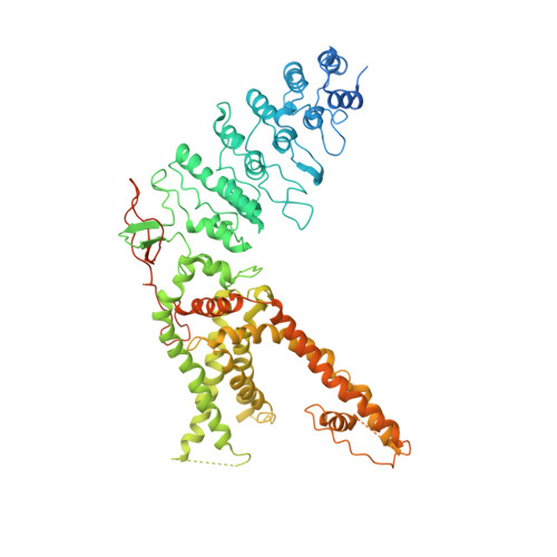
Structure of the full-length TRPV2 channel by cryo-EM.

Huynh, K.W.Cohen, M.R.Jiang, J.Samanta, A.Lodowski, D.T.Zhou, Z.H.Moiseenkova-Bell, V.Y.

(2016) Nat Commun 7: 11130-11130

  • DOI: https://doi.org/10.1038/ncomms11130
  • Primary Citation of Related Structures:  
    5HI9

  • PubMed Abstract: 

    Transient receptor potential (TRP) proteins form a superfamily Ca(2+)-permeable cation channels regulated by a range of chemical and physical stimuli. Structural analysis of a 'minimal' TRP vanilloid subtype 1 (TRPV1) elucidated a mechanism of channel activation by agonists through changes in its outer pore region. Though homologous to TRPV1, other TRPV channels (TRPV2-6) are insensitive to TRPV1 activators including heat and vanilloids. To further understand the structural basis of TRPV channel function, we determined the structure of full-length TRPV2 at ∼5 Å resolution by cryo-electron microscopy. Like TRPV1, TRPV2 contains two constrictions, one each in the pore-forming upper and lower gates. The agonist-free full-length TRPV2 has wider upper and lower gates compared with closed and agonist-activated TRPV1. We propose these newly revealed TRPV2 structural features contribute to diversity of TRPV channels.


  • Organizational Affiliation

    Department of Pharmacology, School of Medicine, Case Western Reserve University, 10900 Euclid Avenue, Wood Building, W151D, Cleveland, Ohio 44106, USA.


Macromolecules
Find similar proteins by:  (by identity cutoff)  |  3D Structure
Entity ID: 1
MoleculeChains Sequence LengthOrganismDetailsImage
Transient Receptor Potential Cation Channel Subfamily V Member 2
A, B, C, D
770Rattus norvegicusMutation(s): 0 
Membrane Entity: Yes 
UniProt
Find proteins for Q9WUD2 (Rattus norvegicus)
Explore Q9WUD2 
Go to UniProtKB:  Q9WUD2
Entity Groups  
Sequence Clusters30% Identity50% Identity70% Identity90% Identity95% Identity100% Identity
UniProt GroupQ9WUD2
Sequence Annotations
Expand
  • Reference Sequence
Experimental Data & Validation

Experimental Data

  • Method: ELECTRON MICROSCOPY
  • Resolution: 4.40 Å
  • Aggregation State: PARTICLE 
  • Reconstruction Method: SINGLE PARTICLE 
EM Software:
TaskSoftware PackageVersion
MODEL REFINEMENTPHENIX1.10.1

Structure Validation

View Full Validation Report


Entry History & Funding Information

Deposition Data


Funding OrganizationLocationGrant Number
American Heart AssociationUnited StatesNCRP Scientist Development Grant 11SDG5280029

Revision History  (Full details and data files)

  • Version 1.0: 2016-03-30
    Type: Initial release
  • Version 1.1: 2016-04-13
    Changes: Database references
  • Version 1.2: 2016-05-11
    Changes: Other
  • Version 1.3: 2017-10-04
    Changes: Author supporting evidence, Data collection
  • Version 1.4: 2024-03-06
    Changes: Data collection, Database references