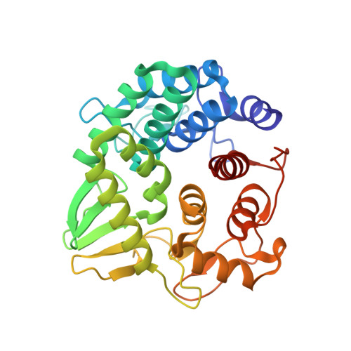
The crystal structure of the endoglucanase Cel10, a family 8 glycosyl hydrolase from Klebsiella pneumoniae
Attigani, A., Sun, L., Wang, Q., Liu, Y., Bai, D., Li, S., Huang, X.(2016) Acta Crystallogr F Struct Biol Commun 72: 870-876
- PubMed: 27917834
- DOI: https://doi.org/10.1107/S2053230X16017891
- Primary Citation of Related Structures:
5GY3 - PubMed Abstract:
Cellulases are produced by microorganisms that grow on cellulose biomass. Here, a cellulase, Cel10, was identified in a strain of Klebsiella pneumoniae isolated from Chinese bamboo rat gut. Analysis of substrate specificity showed that Cel10 is able to hydrolyze amorphous carboxymethyl cellulose (CMC) and crystalline forms of cellulose (Avicel and xylan) but is unable to hydrolyze p-nitrophenol β-D-glucopyranoside (p-NPG), proving that Cel10 is an endoglucanase. A phylogenetic tree analysis indicates that Cel10 belongs to the glycoside hydrolase 8 (GH8) subfamily. In order to further understanding of its substrate specificity, the structure of Cel10 was solved by molecular replacement and refined to 1.76 Å resolution. The overall fold is distinct from those of most other enzymes belonging to the GH8 subfamily. Although it forms the typical (α/α) 6 -barrel motif fold, like Acetobacterxylinum CMCax, one helix is missing. Structural comparisons with Clostridium thermocellum CelA (CtCelA), the best characterized GH8 endoglucanase, revealed that sugar-recognition subsite -3 is completely missing in Cel10. The absence of this subsite correlates to a more open substrate-binding cleft on the cellooligosaccharide reducing-end side.
Organizational Affiliation:
Key Laboratory for Integrated Chinese Traditional and Western Veterinary Medicine and Animal Healthcare, College of Animal Science, Fujian Agriculture and Forestry University, 15 Shang Xia Dian Road, Fuzhou 350002, People's Republic of China.


