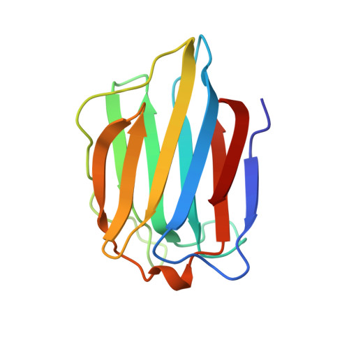
5GAL

CRYSTAL STRUCTURE OF HUMAN GALECTIN-7 IN COMPLEX WITH N-ACETYLLACTOSAMINE


Experimental Data Snapshot

  • Method: X-RAY DIFFRACTION
  • Resolution: 2.00 Å
  • R-Value Free: 0.296 
  • R-Value Work: 0.213 
  • R-Value Observed: 0.213 

wwPDB Validation   3D Report Full Report


This is version 2.2 of the entry. See complete history


Literature

Structural basis for the recognition of carbohydrates by human galectin-7.

Leonidas, D.D.Vatzaki, E.H.Vorum, H.Celis, J.E.Madsen, P.Acharya, K.R.

(1998) Biochemistry 37: 13930-13940

  • DOI: https://doi.org/10.1021/bi981056x
  • Primary Citation of Related Structures:  
    1BKZ, 2GAL, 3GAL, 4GAL, 5GAL

  • PubMed Abstract: 

    Knowledge about carbohydrate recognition domains of galectins, formerly known as S-type animal lectins, is important in understanding their role(s) in cell-cell interactions. Here we report the crystal structure of human galectin-7 (hGal-7), in free form and in the presence of galactose, galactosamine, lactose, and N-acetyl-lactosamine at high resolution. This is the first structure of a galectin determined in both free and carbohydrate-bound forms. The structure shows a fold similar to that of the prototype galectins -1 and -2, but has greater similarity to a related galectin molecule, Gal-10. Even though the carbohydrate-binding residues are conserved, there are significant changes in this pocket due to shortening of a loop structure. The monomeric hGal-7 molecule exists as a dimer in the crystals, but adopts a packing arrangement considerably different from that of Gal-1 and Gal-2, which has implications for carbohydrate recognition.


  • Organizational Affiliation

    Department of Biology and Biochemistry, University of Bath, UK.


Macromolecules
Find similar proteins by:  (by identity cutoff)  |  3D Structure
Entity ID: 1
MoleculeChains Sequence LengthOrganismDetailsImage
GALECTIN-7
A, B
135Homo sapiensMutation(s): 0 
UniProt & NIH Common Fund Data Resources
Find proteins for P47929 (Homo sapiens)
Explore P47929 
Go to UniProtKB:  P47929
PHAROS:  P47929
Entity Groups  
Sequence Clusters30% Identity50% Identity70% Identity90% Identity95% Identity100% Identity
UniProt GroupP47929
Sequence Annotations
Expand
  • Reference Sequence
Oligosaccharides

Help

Entity ID: 2
MoleculeChains Length2D Diagram Glycosylation3D Interactions
beta-D-galactopyranose-(1-4)-2-acetamido-2-deoxy-beta-D-glucopyranose
C
2N/A
Glycosylation Resources
GlyTouCan:  G00055MO
GlyCosmos:  G00055MO
GlyGen:  G00055MO
Experimental Data & Validation

Experimental Data

  • Method: X-RAY DIFFRACTION
  • Resolution: 2.00 Å
  • R-Value Free: 0.296 
  • R-Value Work: 0.213 
  • R-Value Observed: 0.213 
  • Space Group: P 21 21 21
Unit Cell:
Length ( Å )Angle ( ˚ )
a = 54.23α = 90
b = 65.18β = 90
c = 73.39γ = 90
Software Package:
Software NamePurpose
X-PLORmodel building
X-PLORrefinement
DENZOdata reduction
SCALEPACKdata scaling
X-PLORphasing

Structure Validation

View Full Validation Report


Entry History 

Deposition Data

Revision History  (Full details and data files)

  • Version 1.0: 1998-11-04
    Type: Initial release
  • Version 1.1: 2008-03-25
    Changes: Version format compliance
  • Version 1.2: 2011-07-13
    Changes: Non-polymer description, Version format compliance
  • Version 2.0: 2020-07-29
    Type: Remediation
    Reason: Carbohydrate remediation
    Changes: Advisory, Atomic model, Data collection, Derived calculations, Other, Structure summary
  • Version 2.1: 2024-03-13
    Changes: Data collection, Database references, Structure summary
  • Version 2.2: 2024-04-03
    Changes: Refinement description