5FSG

Structure of the hantavirus nucleoprotein provides insights into the mechanism of RNA encapsidation and a template for drug design


Experimental Data Snapshot

  • Method: X-RAY DIFFRACTION
  • Resolution: 3.21 Å
  • R-Value Free: 0.299 
  • R-Value Work: 0.290 
  • R-Value Observed: 0.291 

wwPDB Validation   3D Report Full Report


This is version 3.1 of the entry. See complete history


Literature

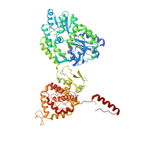
Structure of the Hantavirus Nucleoprotein Provides Insights Into the Mechanism of RNA Encapsidation.

Olal, D.Daumke, O.

(2016) Cell Rep 14: 2092

  • DOI: https://doi.org/10.1016/j.celrep.2016.02.005
  • Primary Citation of Related Structures:  
    5FSG

  • PubMed Abstract: 

    Hantaviruses are etiological agents of life-threatening hemorrhagic fever with renal syndrome and hantavirus cardiopulmonary syndrome. The nucleoprotein (N) of hantavirus is essential for viral transcription and replication, thus representing an attractive target for therapeutic intervention. We have determined the crystal structure of hantavirus N to 3.2 Å resolution. The structure reveals a two-lobed, mostly α-helical structure that is distantly related to that of orthobunyavirus Ns. A basic RNA binding pocket is located at the intersection between the two lobes. We provide evidence that oligomerization is mediated by amino- and C-terminal arms that bind to the adjacent monomers. Based on these findings, we suggest a model for the oligomeric ribonucleoprotein (RNP) complex. Our structure provides mechanistic insights into RNA encapsidation in the genus Hantavirus and constitutes a template for drug discovery efforts aimed at combating hantavirus infections.


  • Organizational Affiliation

    Crystallography, Max Delbrück Center for Molecular Medicine, Robert-Rössle-Strasse 10, 13125 Berlin, Germany. Electronic address: daniel.olal@mdc-berlin.de.


Macromolecules
Find similar proteins by:  (by identity cutoff)  |  3D Structure
Entity ID: 1
MoleculeChains Sequence LengthOrganismDetailsImage
MALTOSE-BINDING PERIPLASMIC PROTEIN, HANTAVIRUS NUCLEOPROTEIN698Hantaan virus 76-118Mutation(s): 0 
UniProt
Find proteins for P0AEX9 (Escherichia coli (strain K12))
Explore P0AEX9 
Go to UniProtKB:  P0AEX9
Find proteins for P05133 (Hantaan virus (strain 76-118))
Explore P05133 
Go to UniProtKB:  P05133
Entity Groups  
Sequence Clusters30% Identity50% Identity70% Identity90% Identity95% Identity100% Identity
UniProt GroupsP05133P0AEX9
Sequence Annotations
Expand
  • Reference Sequence
Oligosaccharides

Help

Entity ID: 2
MoleculeChains Length2D Diagram Glycosylation3D Interactions
alpha-D-glucopyranose-(1-4)-alpha-D-glucopyranose-(1-4)-alpha-D-glucopyranose-(1-4)-alpha-D-glucopyranose
B
4N/A
Glycosylation Resources
GlyTouCan:  G87171PZ
GlyCosmos:  G87171PZ
GlyGen:  G87171PZ
Small Molecules
Modified Residues  1 Unique
IDChains TypeFormula2D DiagramParent
MSE
Query on MSE
A
L-PEPTIDE LINKINGC5 H11 N O2 SeMET
Biologically Interesting Molecules (External Reference) 1 Unique
Experimental Data & Validation

Experimental Data

  • Method: X-RAY DIFFRACTION
  • Resolution: 3.21 Å
  • R-Value Free: 0.299 
  • R-Value Work: 0.290 
  • R-Value Observed: 0.291 
  • Space Group: P 21 21 21
Unit Cell:
Length ( Å )Angle ( ˚ )
a = 69.63α = 90
b = 86.84β = 90
c = 131.5γ = 90
Software Package:
Software NamePurpose
PHENIXrefinement
XDSdata reduction
XDSdata scaling
PHASERphasing

Structure Validation

View Full Validation Report


Entry History 

Deposition Data

Revision History  (Full details and data files)

  • Version 1.0: 2016-04-06
    Type: Initial release
  • Version 2.0: 2019-10-23
    Changes: Atomic model, Data collection, Derived calculations, Other
  • Version 3.0: 2020-07-29
    Type: Remediation
    Reason: Carbohydrate remediation
    Changes: Advisory, Atomic model, Data collection, Derived calculations, Non-polymer description, Structure summary
  • Version 3.1: 2024-01-10
    Changes: Data collection, Database references, Refinement description, Structure summary