5FLU

Structure of a Chaperone-Usher pilus reveals the molecular basis of rod uncoilin


Experimental Data Snapshot

  • Method: ELECTRON MICROSCOPY
  • Resolution: 3.80 Å
  • Aggregation State: FILAMENT 
  • Reconstruction Method: HELICAL 

wwPDB Validation   3D Report Full Report


This is version 1.3 of the entry. See complete history


Literature

Structure of a Chaperone-Usher Pilus Reveals the Molecular Basis of Rod Uncoiling.

Hospenthal, M.K.Redzej, A.Dodson, K.Ukleja, M.Frenz, B.Rodrigues, C.Hultgren, S.J.Dimaio, F.Egelman, E.H.Waksman, G.

(2016) Cell 164: 269

  • DOI: https://doi.org/10.1016/j.cell.2015.11.049
  • Primary Citation of Related Structures:  
    5FLU

  • PubMed Abstract: 

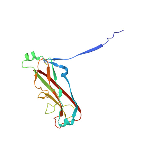
    Types 1 and P pili are prototypical bacterial cell-surface appendages playing essential roles in mediating adhesion of bacteria to the urinary tract. These pili, assembled by the chaperone-usher pathway, are polymers of pilus subunits assembling into two parts: a thin, short tip fibrillum at the top, mounted on a long pilus rod. The rod adopts a helical quaternary structure and is thought to play essential roles: its formation may drive pilus extrusion by preventing backsliding of the nascent growing pilus within the secretion pore; the rod also has striking spring-like properties, being able to uncoil and recoil depending on the intensity of shear forces generated by urine flow. Here, we present an atomic model of the P pilus generated from a 3.8 Å resolution cryo-electron microscopy reconstruction. This structure provides the molecular basis for the rod's remarkable mechanical properties and illuminates its role in pilus secretion.


  • Organizational Affiliation

    Institute of Structural and Molecular Biology, University College London and Birkbeck, Malet Street, London, WC1E 7HX, UK.


Macromolecules
Find similar proteins by:  (by identity cutoff)  |  3D Structure
Entity ID: 1
MoleculeChains Sequence LengthOrganismDetailsImage
PAP FIMBRIAL MAJOR PILIN PROTEIN
A, B, C, D, E
A, B, C, D, E, F, G, H, I, J, K, L, M
163Escherichia coliMutation(s): 0 
UniProt
Find proteins for P04127 (Escherichia coli)
Explore P04127 
Go to UniProtKB:  P04127
Entity Groups  
Sequence Clusters30% Identity50% Identity70% Identity90% Identity95% Identity100% Identity
UniProt GroupP04127
Sequence Annotations
Expand
  • Reference Sequence
Experimental Data & Validation

Experimental Data

  • Method: ELECTRON MICROSCOPY
  • Resolution: 3.80 Å
  • Aggregation State: FILAMENT 
  • Reconstruction Method: HELICAL 
EM Software:
TaskSoftware PackageVersion
RECONSTRUCTIONSPIDER

Structure Validation

View Full Validation Report


Entry History 

Deposition Data

Revision History  (Full details and data files)

  • Version 1.0: 2016-01-13
    Type: Initial release
  • Version 1.1: 2016-01-27
    Changes: Database references
  • Version 1.2: 2018-10-03
    Changes: Author supporting evidence, Data collection
  • Version 1.3: 2019-10-23
    Changes: Data collection, Other