5FJM

Structure of L-Amino acid deaminase from Proteus myxofaciens


Experimental Data Snapshot

  • Method: X-RAY DIFFRACTION
  • Resolution: 2.00 Å
  • R-Value Free: 0.209 
  • R-Value Work: 0.177 
  • R-Value Observed: 0.179 

wwPDB Validation   3D Report Full Report


Ligand Structure Quality Assessment 


This is version 1.2 of the entry. See complete history


Literature

Structure-Function Relationships in L-Amino Acid Deaminase, a Flavoprotein Belonging to a Novel Class of Biotechnologically Relevant Enzymes

Motta, P.Molla, G.Pollegioni, L.Nardini, M.

(2016) J Biol Chem 291: 10457

  • DOI: https://doi.org/10.1074/jbc.M115.703819
  • Primary Citation of Related Structures:  
    5FJM, 5FJN

  • PubMed Abstract: 

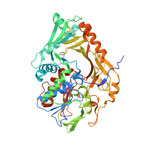
    l-Amino acid deaminase from Proteus myxofaciens (PmaLAAD) is a membrane flavoenzyme that catalyzes the deamination of neutral and aromatic l-amino acids into α-keto acids and ammonia. PmaLAAD does not use dioxygen to re-oxidize reduced FADH2 and thus does not produce hydrogen peroxide; instead, it uses a cytochrome b-like protein as an electron acceptor. Although the overall fold of this enzyme resembles that of known amine or amino acid oxidases, it shows the following specific structural features: an additional novel α+β subdomain placed close to the putative transmembrane α-helix and to the active-site entrance; an FAD isoalloxazine ring exposed to solvent; and a large and accessible active site suitable to bind large hydrophobic substrates. In addition, PmaLAAD requires substrate-induced conformational changes of part of the active site, particularly in Arg-316 and Phe-318, to achieve the correct geometry for catalysis. These studies are expected to pave the way for rationally improving the versatility of this flavoenzyme, which is critical for biocatalysis of enantiomerically pure amino acids.


  • Organizational Affiliation

    From the Dipartimento di Biotecnologie e Scienze della Vita, Università degli Studi deII'Insubria, via J. H. Dunant 3, 21100 Varese.


Macromolecules
Find similar proteins by:  (by identity cutoff)  |  3D Structure
Entity ID: 1
MoleculeChains Sequence LengthOrganismDetailsImage
L-AMINO ACID DEAMINASE
A, B
456Proteus myxofaciensMutation(s): 0 
UniProt
Find proteins for A0A158RFS7 (Proteus myxofaciens)
Explore A0A158RFS7 
Go to UniProtKB:  A0A158RFS7
Entity Groups  
Sequence Clusters30% Identity50% Identity70% Identity90% Identity95% Identity100% Identity
UniProt GroupA0A158RFS7
Sequence Annotations
Expand
  • Reference Sequence
Small Molecules
Ligands 1 Unique
IDChains Name / Formula / InChI Key2D Diagram3D Interactions
FAD
Query on FAD

Download Ideal Coordinates CCD File 
C [auth A],
D [auth B]
FLAVIN-ADENINE DINUCLEOTIDE
C27 H33 N9 O15 P2
VWWQXMAJTJZDQX-UYBVJOGSSA-N
Experimental Data & Validation

Experimental Data

  • Method: X-RAY DIFFRACTION
  • Resolution: 2.00 Å
  • R-Value Free: 0.209 
  • R-Value Work: 0.177 
  • R-Value Observed: 0.179 
  • Space Group: P 1 21 1
Unit Cell:
Length ( Å )Angle ( ˚ )
a = 72.76α = 90
b = 93.71β = 102.85
c = 74.69γ = 90
Software Package:
Software NamePurpose
REFMACrefinement
XDSdata reduction
SCALAdata scaling
PHASERphasing

Structure Validation

View Full Validation Report


Ligand Structure Quality Assessment 


Entry History 

Deposition Data

Revision History  (Full details and data files)

  • Version 1.0: 2016-04-06
    Type: Initial release
  • Version 1.1: 2016-06-15
    Changes: Database references
  • Version 1.2: 2024-01-10
    Changes: Data collection, Database references, Derived calculations, Other, Refinement description