5FD0

Streptomyces plicatus N-acetyl-beta-hexosaminidase in complex with NAGlucal


Experimental Data Snapshot

  • Method: X-RAY DIFFRACTION
  • Resolution: 2.00 Å
  • R-Value Free: 0.180 
  • R-Value Work: 0.150 
  • R-Value Observed: 0.151 

wwPDB Validation   3D Report Full Report


Ligand Structure Quality Assessment 


This is version 1.3 of the entry. See complete history


Literature

N-Acetyl glycals are tight-binding and environmentally insensitive inhibitors of hexosaminidases.

Santana, A.G.Vadlamani, G.Mark, B.L.Withers, S.G.

(2016) Chem Commun (Camb) 52: 7943-7946

  • DOI: https://doi.org/10.1039/c6cc02520j
  • Primary Citation of Related Structures:  
    5FCZ, 5FD0

  • PubMed Abstract: 

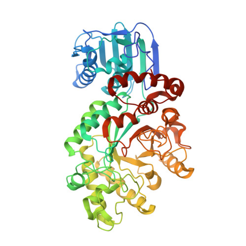
    Mono-, di- and trisaccharide derivatives of 1,2-unsaturated N-acetyl-d-glucal have been synthesized and shown to function as tight-binding inhibitors/slow substrates of representative hexosaminidases. Turnover is slow and not observed in the thioamide analogue, allowing determination of the 3-dimensional structure of the complex. Inhibition is insensitive to pH and to mutation of key catalytic residues, consistent with the uncharged character of the inhibitor. These properties could render this inhibitor class less prone to development of resistance.


  • Organizational Affiliation

    Department of Chemistry, University of British Columbia, 2036 Main Mall, Vancouver, British Columbia V6T1Z3, Canada. withers@chem.ubc.ca.


Macromolecules
Find similar proteins by:  (by identity cutoff)  |  3D Structure
Entity ID: 1
MoleculeChains Sequence LengthOrganismDetailsImage
B-N-acetylhexosaminidase512Streptomyces plicatusMutation(s): 0 
Gene Names: hex
UniProt
Find proteins for O85361 (Streptomyces plicatus)
Explore O85361 
Go to UniProtKB:  O85361
Entity Groups  
Sequence Clusters30% Identity50% Identity70% Identity90% Identity95% Identity100% Identity
UniProt GroupO85361
Sequence Annotations
Expand
  • Reference Sequence
Small Molecules
Binding Affinity Annotations 
IDSourceBinding Affinity
NAG Binding MOAD:  5FD0 Ki: 2400 (nM) from 1 assay(s)
Experimental Data & Validation

Experimental Data

  • Method: X-RAY DIFFRACTION
  • Resolution: 2.00 Å
  • R-Value Free: 0.180 
  • R-Value Work: 0.150 
  • R-Value Observed: 0.151 
  • Space Group: P 61 2 2
Unit Cell:
Length ( Å )Angle ( ˚ )
a = 133.17α = 90
b = 133.17β = 90
c = 176.525γ = 120
Software Package:
Software NamePurpose
PHENIXrefinement
Aimlessdata scaling
PHASERphasing
CrystalCleardata collection

Structure Validation

View Full Validation Report


Ligand Structure Quality Assessment 


Entry History & Funding Information

Deposition Data


Funding OrganizationLocationGrant Number
Cystic Fibrosis CanadaCanada--

Revision History  (Full details and data files)

  • Version 1.0: 2016-10-26
    Type: Initial release
  • Version 1.1: 2017-11-01
    Changes: Author supporting evidence, Derived calculations
  • Version 1.2: 2020-07-29
    Type: Remediation
    Reason: Carbohydrate remediation
    Changes: Data collection, Derived calculations, Structure summary
  • Version 1.3: 2023-09-27
    Changes: Data collection, Database references, Refinement description, Structure summary