5EFR

Crystal Structure of a BamA-BamD fusion


Experimental Data Snapshot

  • Method: X-RAY DIFFRACTION
  • Resolution: 2.00 Å
  • R-Value Free: 0.232 
  • R-Value Work: 0.211 
  • R-Value Observed: 0.212 

wwPDB Validation   3D Report Full Report


This is version 1.4 of the entry. See complete history


Literature

The Structure of a BamA-BamD Fusion Illuminates the Architecture of the beta-Barrel Assembly Machine Core.

Bergal, H.T.Hopkins, A.H.Metzner, S.I.Sousa, M.C.

(2016) Structure 24: 243-251

  • DOI: https://doi.org/10.1016/j.str.2015.10.030
  • Primary Citation of Related Structures:  
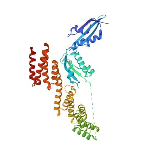
    5EFR

  • PubMed Abstract: 

    The β-barrel assembly machine (BAM) mediates folding and insertion of integral β-barrel outer membrane proteins (OMPs) in Gram-negative bacteria. Of the five BAM subunits, only BamA and BamD are essential for cell viability. Here we present the crystal structure of a fusion between BamA POTRA4-5 and BamD from Rhodothermus marinus. The POTRA5 domain binds BamD between its tetratricopeptide repeats 3 and 4. The interface structural elements are conserved in the Escherichia coli proteins, which allowed structure validation by mutagenesis and disulfide crosslinking in E. coli. Furthermore, the interface is consistent with previously reported mutations that impair BamA-BamD binding. The structure serves as a linchpin to generate a BAM model where POTRA domains and BamD form an elongated periplasmic ring adjacent to the membrane with a central cavity approximately 30 × 60 Å wide. We propose that nascent OMPs bind this periplasmic ring prior to insertion and folding by BAM.


  • Organizational Affiliation

    Department of Chemistry and Biochemistry, University of Colorado at Boulder, Boulder, CO 80309, USA.


Macromolecules
Find similar proteins by:  (by identity cutoff)  |  3D Structure
Entity ID: 1
MoleculeChains Sequence LengthOrganismDetailsImage
BamA-BamD fusion Protein446Rhodothermus marinusMutation(s): 0 
Entity Groups  
Sequence Clusters30% Identity50% Identity70% Identity90% Identity95% Identity100% Identity
Sequence Annotations
Expand
  • Reference Sequence
Experimental Data & Validation

Experimental Data

  • Method: X-RAY DIFFRACTION
  • Resolution: 2.00 Å
  • R-Value Free: 0.232 
  • R-Value Work: 0.211 
  • R-Value Observed: 0.212 
  • Space Group: P 1 21 1
Unit Cell:
Length ( Å )Angle ( ˚ )
a = 69.909α = 90
b = 48.062β = 104.31
c = 78.027γ = 90
Software Package:
Software NamePurpose
HKL-2000data scaling
PHENIXrefinement
PDB_EXTRACTdata extraction
HKL-2000data reduction
PHENIXphasing

Structure Validation

View Full Validation Report


Entry History & Funding Information

Deposition Data


Funding OrganizationLocationGrant Number
National Institutes of Health/National Institute Of Allergy and Infectious Diseases (NIH/NIAID)United StatesAI080709

Revision History  (Full details and data files)

  • Version 1.0: 2016-01-27
    Type: Initial release
  • Version 1.1: 2016-02-10
    Changes: Database references
  • Version 1.2: 2017-09-27
    Changes: Author supporting evidence, Derived calculations
  • Version 1.3: 2019-12-11
    Changes: Author supporting evidence
  • Version 1.4: 2023-09-27
    Changes: Data collection, Database references, Refinement description