5DUY

Structure of lectin from the sea mussel Crenomytilus grayanus


Experimental Data Snapshot

  • Method: X-RAY DIFFRACTION
  • Resolution: 2.12 Å
  • R-Value Free: 0.184 
  • R-Value Work: 0.139 
  • R-Value Observed: 0.141 

wwPDB Validation   3D Report Full Report


This is version 1.4 of the entry. See complete history


Literature

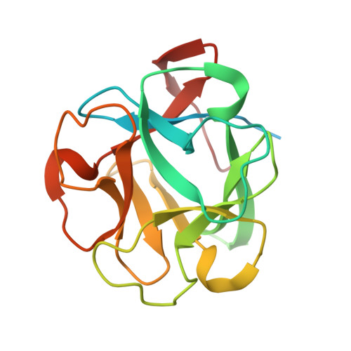
Structure of a lectin from the sea mussel Crenomytilus grayanus (CGL).

Jakob, M.Lubkowski, J.O'Keefe, B.R.Wlodawer, A.

(2015) Acta Crystallogr F Struct Biol Commun 71: 1429-1436

  • DOI: https://doi.org/10.1107/S2053230X15019858
  • Primary Citation of Related Structures:  
    5DUY

  • PubMed Abstract: 

    CGL is a 150 amino-acid residue lectin that was originally isolated from the sea mussel Crenomytilus grayanus. It is specific for binding GalNAc/Gal-containing carbohydrate moieties and in general does not share sequence homology with other known galectins or lectins. Since CGL displays antibacterial, antifungal and antiviral activities, and interacts with high affinity with mucin-type receptors, which are abundant on some cancer cells, knowledge of its structure is of significant interest. Conditions have been established for the expression, purification and crystallization of a recombinant variant of CGL. The crystal structure of recombinant CGL was determined and refined at a resolution of 2.12 Å. The amino-acid sequence of CGL contains three homologous regions (73% similarity) and the folded protein has a β-trefoil topology. Structural comparison of CGL with the closely related lectin MytiLec allowed description of the glycan-binding pockets.


  • Organizational Affiliation

    Macromolecular Crystallography Laboratory, Center for Cancer Research, National Cancer Institute at Frederick, Frederick, MD 21702-1201, USA.


Macromolecules
Find similar proteins by:  (by identity cutoff)  |  3D Structure
Entity ID: 1
MoleculeChains Sequence LengthOrganismDetailsImage
GalNAc/Gal-specific lectin
A, B, C, D, E
A, B, C, D, E, F
181Crenomytilus grayanusMutation(s): 0 
UniProt
Find proteins for H2FH31 (Crenomytilus grayanus)
Explore H2FH31 
Go to UniProtKB:  H2FH31
Entity Groups  
Sequence Clusters30% Identity50% Identity70% Identity90% Identity95% Identity100% Identity
UniProt GroupH2FH31
Sequence Annotations
Expand
  • Reference Sequence
Small Molecules
Ligands 1 Unique
IDChains Name / Formula / InChI Key2D Diagram3D Interactions
GOL
Query on GOL

Download Ideal Coordinates CCD File 
G [auth A]
H [auth A]
I [auth A]
J [auth B]
K [auth B]
G [auth A],
H [auth A],
I [auth A],
J [auth B],
K [auth B],
L [auth B],
M [auth C],
N [auth C],
O [auth C],
P [auth D],
Q [auth D],
R [auth D],
S [auth E],
T [auth E],
U [auth E],
V [auth F],
W [auth F],
X [auth F]
GLYCEROL
C3 H8 O3
PEDCQBHIVMGVHV-UHFFFAOYSA-N
Experimental Data & Validation

Experimental Data

  • Method: X-RAY DIFFRACTION
  • Resolution: 2.12 Å
  • R-Value Free: 0.184 
  • R-Value Work: 0.139 
  • R-Value Observed: 0.141 
  • Space Group: C 1 2 1
Unit Cell:
Length ( Å )Angle ( ˚ )
a = 137.289α = 90
b = 68.337β = 110.55
c = 131.694γ = 90
Software Package:
Software NamePurpose
REFMACrefinement
HKL-2000data reduction
HKL-2000data scaling
PHASERphasing
MR-Rosettaphasing

Structure Validation

View Full Validation Report


Entry History 

Deposition Data

Revision History  (Full details and data files)

  • Version 1.0: 2015-11-11
    Type: Initial release
  • Version 1.1: 2015-11-18
    Changes: Database references
  • Version 1.2: 2015-12-23
    Changes: Structure summary
  • Version 1.3: 2017-11-01
    Changes: Author supporting evidence, Derived calculations
  • Version 1.4: 2024-03-06
    Changes: Data collection, Database references