5DPN

Engineered CBM X-2 L110F in complex with branched carbohydrate XXXG.


Experimental Data Snapshot

  • Method: X-RAY DIFFRACTION
  • Resolution: 1.60 Å
  • R-Value Free: 0.250 
  • R-Value Work: 0.223 
  • R-Value Observed: 0.224 

  • Method: NEUTRON DIFFRACTION
  • Resolution: 1.61 Å
  • R-Value Free: 0.204 
  • R-Value Work: 0.163 
  • R-Value Observed: 0.165 

wwPDB Validation   3D Report Full Report


This is version 2.0 of the entry. See complete history


Literature

Neutron Crystallographic Studies Reveal Hydrogen Bond and Water-Mediated Interactions between a Carbohydrate-Binding Module and Its Bound Carbohydrate Ligand.

Fisher, S.Z.von Schantz, L.Hakansson, M.Logan, D.T.Ohlin, M.

(2015) Biochemistry 54: 6435-6438

  • DOI: https://doi.org/10.1021/acs.biochem.5b01058
  • Primary Citation of Related Structures:  
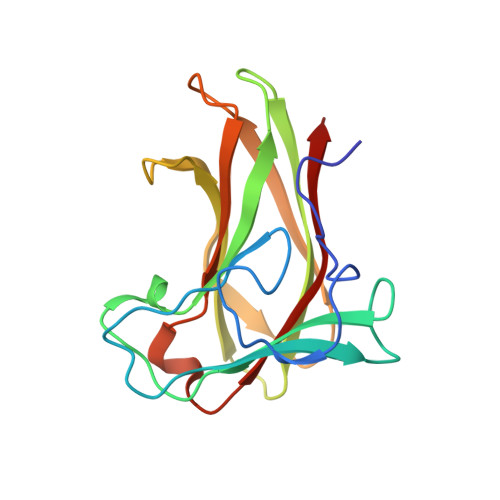
    5DPN

  • PubMed Abstract: 

    Carbohydrate-binding modules (CBMs) are key components of many carbohydrate-modifying enzymes. CBMs affect the activity of these enzymes by modulating bonding and catalysis. To further characterize and study CBM-ligand binding interactions, neutron crystallographic studies of an engineered family 4-type CBM in complex with a branched xyloglucan ligand were conducted. The first neutron crystal structure of a CBM-ligand complex reported here shows numerous atomic details of hydrogen bonding and water-mediated interactions and reveals the charged state of key binding cleft amino acid side chains.


  • Organizational Affiliation

    European Spallation Source , S-221 00 Lund, Sweden.


Macromolecules
Find similar proteins by:  (by identity cutoff)  |  3D Structure
Entity ID: 1
MoleculeChains Sequence LengthOrganismDetailsImage
Xylanase167Rhodothermus marinusMutation(s): 7 
UniProt
Find proteins for Q7WTN6 (Rhodothermus marinus)
Explore Q7WTN6 
Go to UniProtKB:  Q7WTN6
Entity Groups  
Sequence Clusters30% Identity50% Identity70% Identity90% Identity95% Identity100% Identity
UniProt GroupQ7WTN6
Sequence Annotations
Expand
  • Reference Sequence
Oligosaccharides

Help

Entity ID: 2
MoleculeChains Length2D Diagram Glycosylation3D Interactions
alpha-D-xylopyranose-(1-6)-beta-D-glucopyranose-(1-4)-[alpha-D-xylopyranose-(1-6)]beta-D-glucopyranose-(1-4)-[alpha-D-xylopyranose-(1-6)]beta-D-glucopyranose-(1-4)-beta-D-glucopyranose
B
7N/A
Glycosylation Resources
GlyTouCan:  G24181CY
GlyCosmos:  G24181CY
Small Molecules
Ligands 1 Unique
IDChains Name / Formula / InChI Key2D Diagram3D Interactions
CA
Query on CA

Download Ideal Coordinates CCD File 
C [auth A]CALCIUM ION
Ca
BHPQYMZQTOCNFJ-UHFFFAOYSA-N
Experimental Data & Validation

Experimental Data

  • Method: X-RAY DIFFRACTION
  • Resolution: 1.60 Å
  • R-Value Free: 0.250 
  • R-Value Work: 0.223 
  • R-Value Observed: 0.224 
  • Space Group: P 21 21 2
  • Method: NEUTRON DIFFRACTION
  • Resolution: 1.61 Å
  • R-Value Free: 0.204 
  • R-Value Work: 0.163 
  • R-Value Observed: 0.165 
  • Space Group: P 21 21 2
Unit Cell:
Length ( Å )Angle ( ˚ )
a = 72.977α = 90
b = 49.856β = 90
c = 45.091γ = 90
Software Package:
Software NamePurpose
PHENIXrefinement
DENZOdata reduction
SCALEPACKdata scaling
Cootmodel building

Structure Validation

View Full Validation Report


Entry History 

Deposition Data

  • Released Date: 2015-10-28 
  • Deposition Author(s): Ohlin, M.

Revision History  (Full details and data files)

  • Version 1.0: 2015-10-28
    Type: Initial release
  • Version 1.1: 2015-11-04
    Changes: Database references
  • Version 1.2: 2018-01-17
    Changes: Data collection
  • Version 1.3: 2018-02-28
    Changes: Data collection
  • Version 1.4: 2018-03-07
    Changes: Data collection
  • Version 2.0: 2020-07-29
    Type: Remediation
    Reason: Carbohydrate remediation
    Changes: Advisory, Atomic model, Data collection, Derived calculations, Structure summary