5DOR

P2 Integrase catalytic domain in space group P21


Experimental Data Snapshot

  • Method: X-RAY DIFFRACTION
  • Resolution: 2.50 Å
  • R-Value Free: 0.275 
  • R-Value Work: 0.230 
  • R-Value Observed: 0.232 

wwPDB Validation   3D Report Full Report


This is version 1.1 of the entry. See complete history


Literature

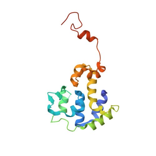
Crystal structure of the bacteriophage P2 integrase catalytic domain.

Skaar, K.Claesson, M.Odegrip, R.Hogbom, M.Haggard-Ljungquist, E.Stenmark, P.

(2015) FEBS Lett 589: 3556-3563

  • DOI: https://doi.org/10.1016/j.febslet.2015.09.026
  • Primary Citation of Related Structures:  
    5C6K, 5DOR

  • PubMed Abstract: 

    Bacteriophage P2 is a temperate phage capable of integrating its DNA into the host genome by site-specific recombination upon lysogenization. Integration and excision of the phage genome requires P2 integrase, which performs recognition, cleavage and joining of DNA during these processes. This work presents the high-resolution crystal structure of the catalytic domain of P2 integrase, and analysis of the structure-function relationship of several previously identified non-functional P2 integrase mutants. The DNA binding area is characterized by a large positively charged patch, harboring key residues. The structure reveals potential for large dimer flexibility, likely essential for rearrangement of DNA strands upon integration and excision of the phage DNA.


  • Organizational Affiliation

    Department of Biochemistry and Biophysics, Stockholm University, Stockholm, Sweden.


Macromolecules
Find similar proteins by:  (by identity cutoff)  |  3D Structure
Entity ID: 1
MoleculeChains Sequence LengthOrganismDetailsImage
Integrase
A, B, C, D
176Peduovirus P2Mutation(s): 0 
Gene Names: int
UniProt
Find proteins for P36932 (Escherichia phage P2)
Explore P36932 
Go to UniProtKB:  P36932
Entity Groups  
Sequence Clusters30% Identity50% Identity70% Identity90% Identity95% Identity100% Identity
UniProt GroupP36932
Sequence Annotations
Expand
  • Reference Sequence
Experimental Data & Validation

Experimental Data

  • Method: X-RAY DIFFRACTION
  • Resolution: 2.50 Å
  • R-Value Free: 0.275 
  • R-Value Work: 0.230 
  • R-Value Observed: 0.232 
  • Space Group: P 1 21 1
Unit Cell:
Length ( Å )Angle ( ˚ )
a = 64.11α = 90
b = 92.225β = 96.34
c = 64.937γ = 90
Software Package:
Software NamePurpose
REFMACrefinement
XDSdata reduction
Aimlessdata scaling
MOLREPphasing

Structure Validation

View Full Validation Report


Entry History & Funding Information

Deposition Data


Funding OrganizationLocationGrant Number
Swedish Research CouncilSweden2010-5200, 2014-5667
Wenner-Gren FoundationSweden--
Carl Tryggers FoundationSweden--

Revision History  (Full details and data files)

  • Version 1.0: 2015-12-02
    Type: Initial release
  • Version 1.1: 2024-01-10
    Changes: Data collection, Database references, Refinement description